Chondrometrics


Chondrometrics is a leading provider of medical image analysis services to researchers in academia and in the pharmaceutical industry

ABOUT US

ABOUT CHONDROMETRICS GMBH

Segmented Knee Joint

Chondrometrics was founded in 2003 and is located at Freilassing, Germany, next to the Austrian border; it is a leading provider of medical image analysis for academia and industry. Using a regulatory compliant analysis pipeline, the company relies on MRI to offer quantitative imaging analysis of articular tissues and structural pathology, for research, epidemiological, and interventional studies, Its 20-year experience in high quality image segmentation has recently been supplemented with fully automated image analysis approaches, using deep-learning (AI) technology. The ultimate goal is to qualify imaging endpoints as surrogate markers of clinical outcome in clinical trials, mainly for the regulatory approval of disease-modifying osteoarthritis drugs (DMOADs).

Where We Come From - Where We Are
Chondrometrics GmbH has been founded by Felix Eckstein and Reinhard Putz, as a spin-off from the Ludwig-Maximilians Universität München (Munich, Germany). In 2004, the company transitioned to Ainring, in the Southeast Germany, and in 2021 to its current location at Ludwig-Zeller-Str. 12, 83395 Freilassing, Germany, next to the border of Salzburg, Austria.

What We Do
Chondrometrics provides medical image analysis services to researchers in academia as well as in the medical device and pharmaceutical industry. The company relies on magnetic resonance imaging (MRI) to quantitatively analyze synovial joint structure and pathology, including (but not limited to) cartilage morphology and relaxometry (e.g., T2), meniscus morphology and relative position, bone size and shape, muscle and adipose tissue mass, synovitis, and properties of other musculoskeletal tissues. We have developed a highly efficient software platform and rely on a team of well-trained readers with >20 years of experience in manual segmentations of MRIs, and the Chondrometrics 3.0 software and reading process comply with current regulatory requirements. The company maintains a GCP-compliant quality management system that is certified according to  EN ISO 13485:2021 by medical device certification GmbH (MDC) and is regularly audited MDC and other partners and clients. The software platform used represents a Class-IIa medical device product according to regulation (EU) 2017/745 (Annex IX). Chondrometrics recently started segmentation and analysis services using novel, fully automated technology using deep-learning (AI) approaches, specifically convolutional neural networks (CNNs). In this area, the company profits from a rich resource of tissues segmentations generated over the years by its expert readers that can be used for training and validation of versatile and specific models for automated analysis in various contexts.

Why We Do It
We use quantitative imaging measures to identify different “morpho-types” of human joints that may entail certain risk profiles and thus require adapted therapies for improved disease outcome. These endpoint also help investigating male-female differences in articular tissue anatomy and pathology, helping to better understand and appreciate sex-differences in osteoarthritis in general. They can further elucidate resilience towards joint disease through unraveling functional adaptation processes of articular tissues. Imaging endpoints in epidemiological studies can identify risk factors of incidence and progression in osteoarthritis. Eventually, these quantitative meassures may be qualified as surrogate markers for clinical outcome (measures of how a patient fees, functions, or [her/his joint] survives) in clinical trials, mainly for the regulatory approval of disease-modifying osteoarthritis drugs (DMOADs).

What’s the Problem
Osteoarthritis is a leading cause of functional disability worldwide, affecting millions of people. This devastating degenerative joint disease causes structural changes in joint tissues, leading to pain, stiffness, and functional disability. Early diagnosis and careful clinical management are crucial to maintaining the patients' quality of life. With an aging population, the prevalence of osteoarthritis is bound to rise, stressing the need for effective and sustainable therapy. Apart from lifestyle changes and physiotherapy, current medical intervention is limited to managing pain. No medication is currently available to modify the structural pathology of articular tissues, disease-progression, and the natural history of osteoarthritis.

Where We Are Heading
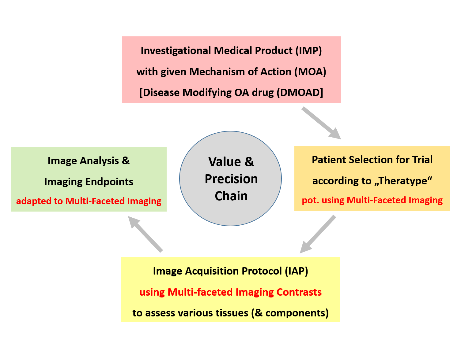
The quantitative imaging measures developed and provided by Chondrometrics may be used in proof-of-concept (POC) trials of structural efficacy and safety. These may ensure that the intended mode-of-action (MOA) of a DMOAD targets the designated tissue, that the structural treatment effect is of sufficient magnitude to impact long-term clinical outcome, and that a potential sudden relieve in pain has no deleterious effect on the articular tissues. In larger follow-up studies, our methodologies can help demonstrating whether the drug's impact on long term clinical benefit (for instance in reducing knee replacements rates) is indeed related to the assumed MOA and target tissue, so that the putative DMOAD can obtain either full or preliminary/conditional approval for its use in osteoarthritis patients. By being validated, reliable, precise, and highly sensitive to both change and interventions, Chondrometrics’ technology helps to make clinical trials more efficient, i.e. more streamlined, faster, and more affordable. Providing expert consulting based on over 30 years of academic experience, we can ensure that the image acquisition protocol of the study will effectively support the desired imaging measures, that the imaging measures selected will match to the drug’s unique molecular profile, and that patient selection criteria match both with the drug’s MOA and the selected imaging endpoints. This type of alignment maximizes the chances of a clinical trial being successful, in that it provides clear and unambiguous answers as to whether an investigational medical product (IMP) has relevant impact on the structural pathology of joint tissues and on the natural progression of the disease (osteoarthritis).

Interested to learn more and here about the research done by company representatives.
Please visit the RESEARCH Section on this page. Contact: eckstein@chondrometrics.de
NEWS

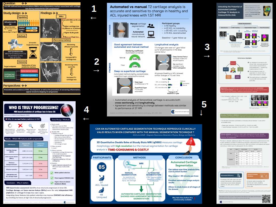
FIRST TWO ORIGINAL ARTICLES PUBLISHED ON AUTOMATED ANAYLSIS OF CARTILAGE T2 (OA-BIO)

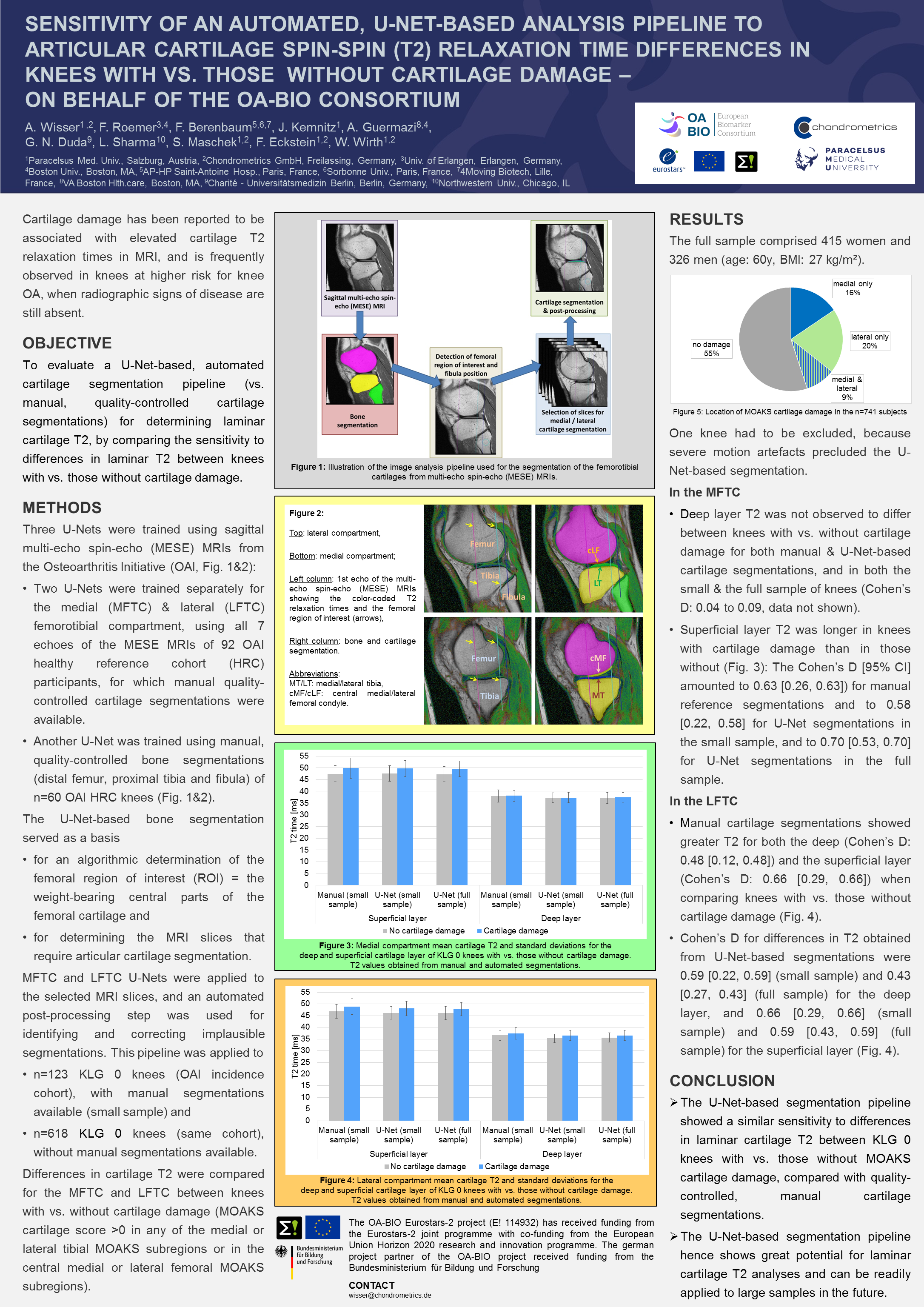
Technical and clinical validation of deep-learning based analysis of laminar properties of knee articular cartilage in two models of early osteoarthritis 

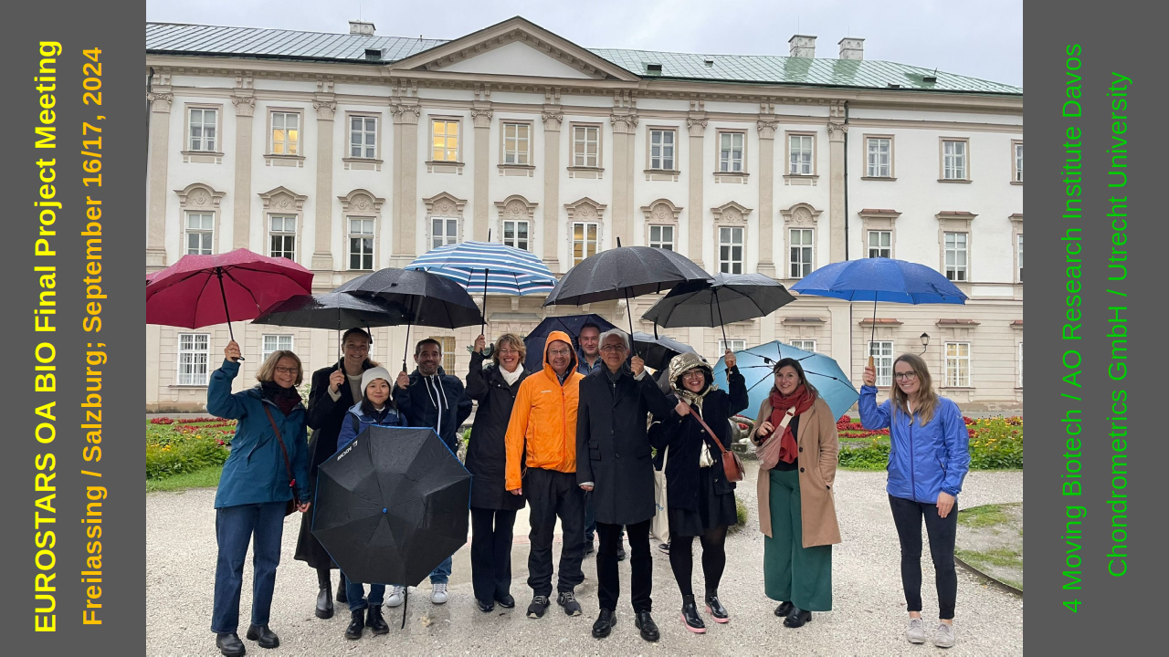
We are proud to announce publication of our two first full original papers generated from OA-BIO, a €4.2 Million Eurostars grant awarded to a consortium of 4 partners: 4Moving Biotech, AO Research Institute Davos (ARI), Utrecht University, and Chondrometrics GmbH. The four-year project aims at developing life-changing therapy for osteoarthritis, using a biomarker-driven approach

The two papers published have just appeared in Skeletal Radiology and in Quantitative Imaging in Medicine and Surgery, two well-established journals in the field of Musculoskeletal Imaging:

  1. Evaluation of an automated laminar cartilage T2 relaxation time analysis method in an early osteoarthritis model by Wolfgang Wirth et al.
    Skeletal Radiol 2024 Sep 4. Online ahead of print.
  2. Clinical validation of fully automated laminar knee cartilage transverse relaxation time (T2) analysis in anterior cruciate ligament (ACL)-injured knees, by Felix Eckstein et al.
    Quantitative Imaging in Medicine and Surgery (QIMS) 2024: 14: 4319-4332.

These make use of two (human) models of early osteoarthritis that were already outlined in our 2020 "Year in review" (imaging) article in Osteoarthritis & Cartilage

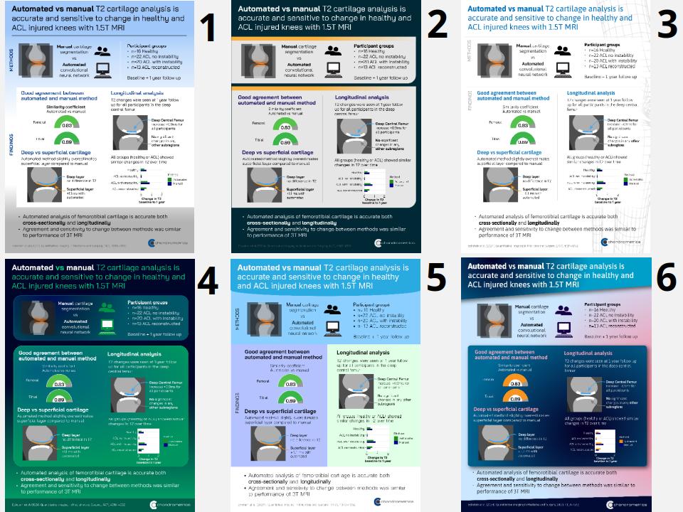
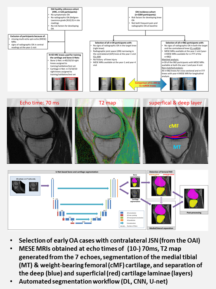
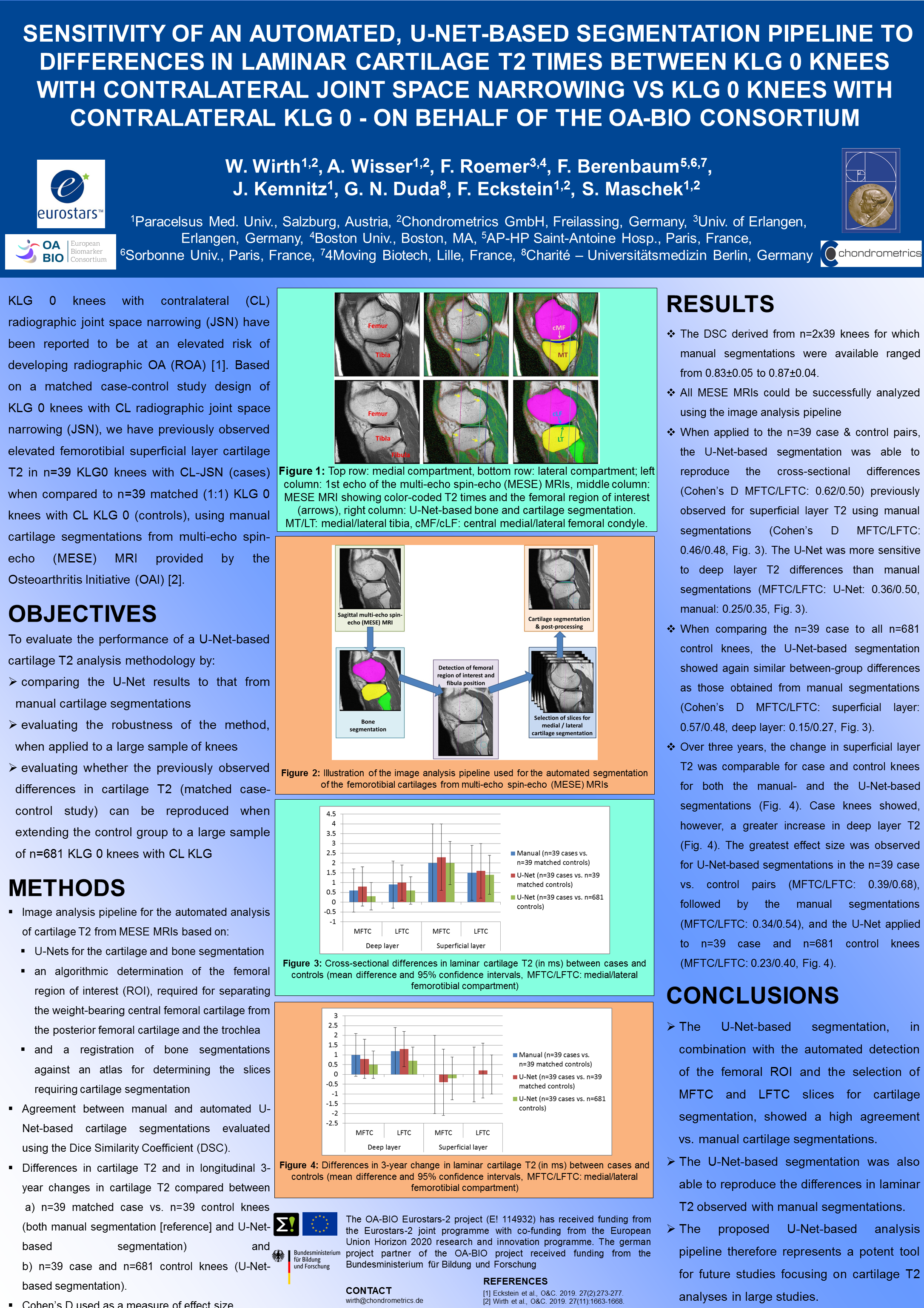
The first publication relies on target knees withOUT radiographic osteoartritis, but with contra-lateral knee radiographic joint space narrowing (JSN); the second one on patients with recent (anterior) cruciate ligament (ACL) injury. The overarching purpose was to technically and clinically validate an automated approach of cartilage MRI transverserelaxation time (T2) analysis.

In the two above papers we demonstrate that:

  1. Cartilage segmentation by deep-learning (CNN- and U net-) based methodology applied to multiple echo spin echo (MESE) MRIs shows high agreement (DSCs) with expert reader segmentation
  2. Automated analysis of T2 in the deep and superficialcartilage layer is accurate when compared with that derived from manual segmentations
  3. Sensitivity to clinically important T2 differences or longitudinal T2 changes is similar between the automated vs. the traditional (expert manual segmentation) approach.

The automated analysis method is currently extended to its application in a quantitative dual echo steady state (qDESS sequence), a more efficient, high-resolution and rapid-acquisition 3D-MRI method for evaluating cartilage T2 (Paper). This ongoing work was recently presented at the International Workshop of Osteoarthritis Imaging (IWOAI) in Marrakech, Morocco, and it is currently submitted for full publication as an original paper.

We thank our co-authors: Susanne Maschek, Anna Wisser, Jana Eder, Christian Baumgartner, Akshay Chaudhari, Francis Berenbaum, Nicholas Brisson, and Georg Duda, and we thank the Chondrometrics expert readers for diligent cartilage segmentation: Jana Daimer, Gudrun Goldmann, Sabine Mühlsimer, Annette Thebis, and Barbara Wehr.

The MRI image analysis in the above studies was funded through OA BIO (E! 114932; Eurostars 2/ Horizon 2020) that supports early clinical development of 4P004, a putative first-in-class disease modifying osteoarthritis drug (DMOAD), as well as validation of osteoarthritis biomarkers required in its clinical development.

Automated Analysis of Cartilage


SPECIAL ISSUE REVIEW ARTICLES
OSTEOARTHRITIS & CARTILAGE (OAC) and OAC OPEN

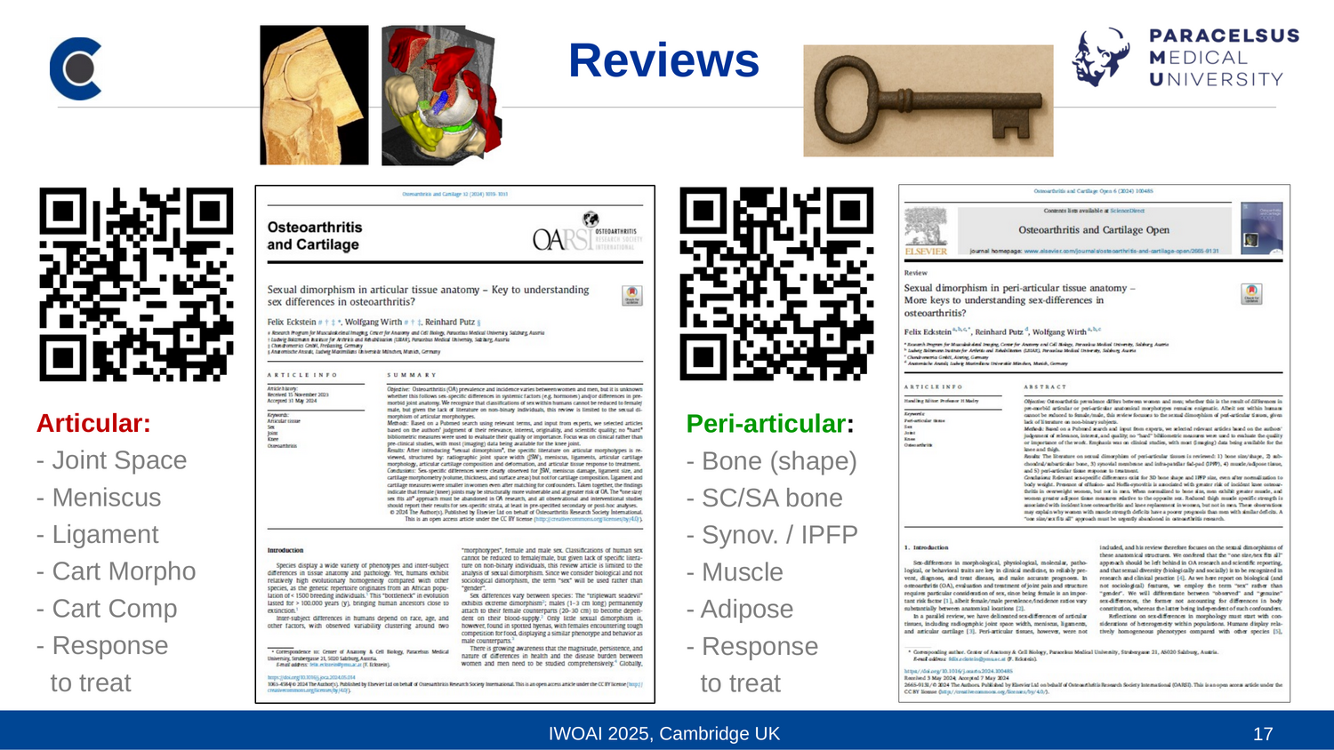
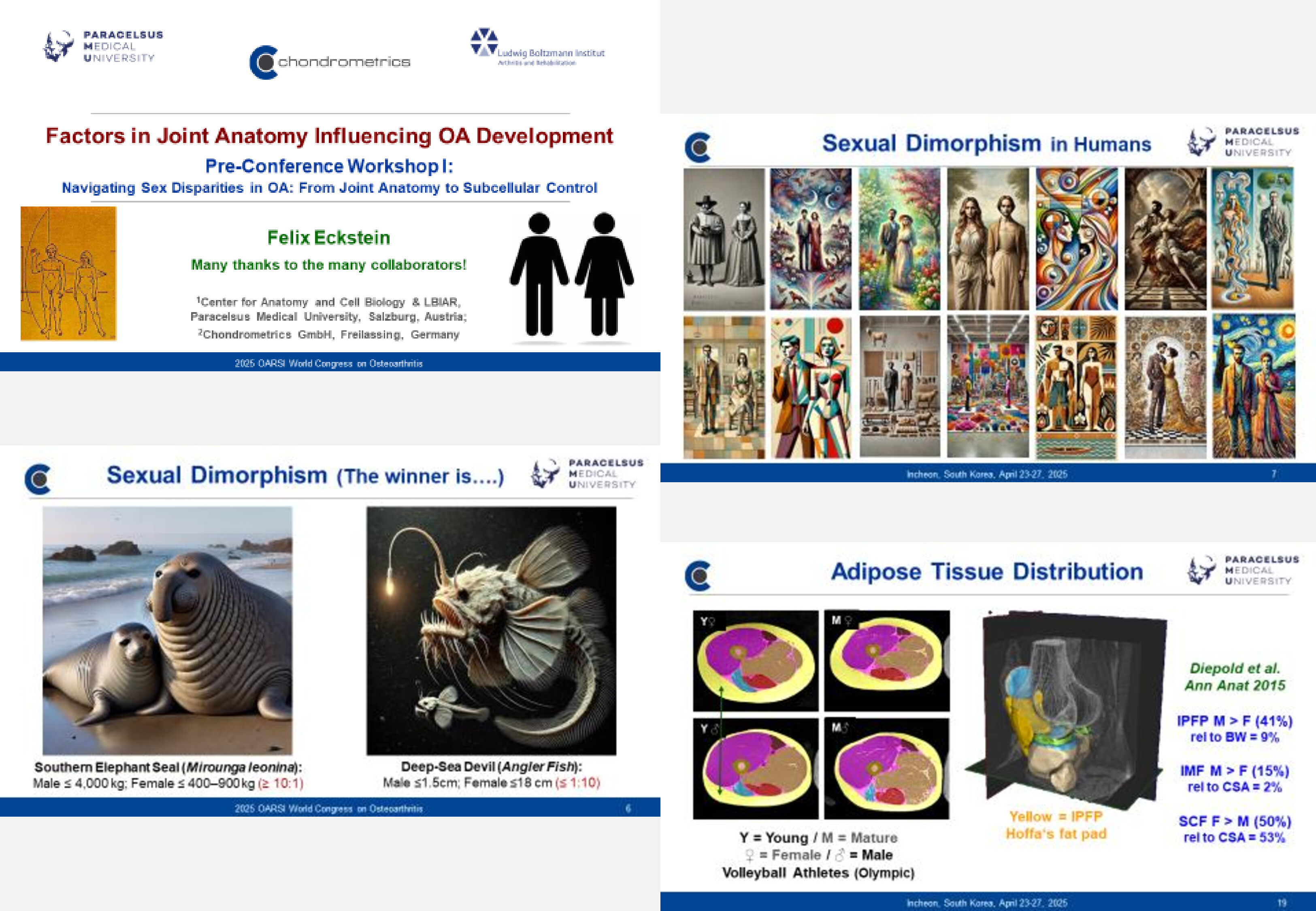
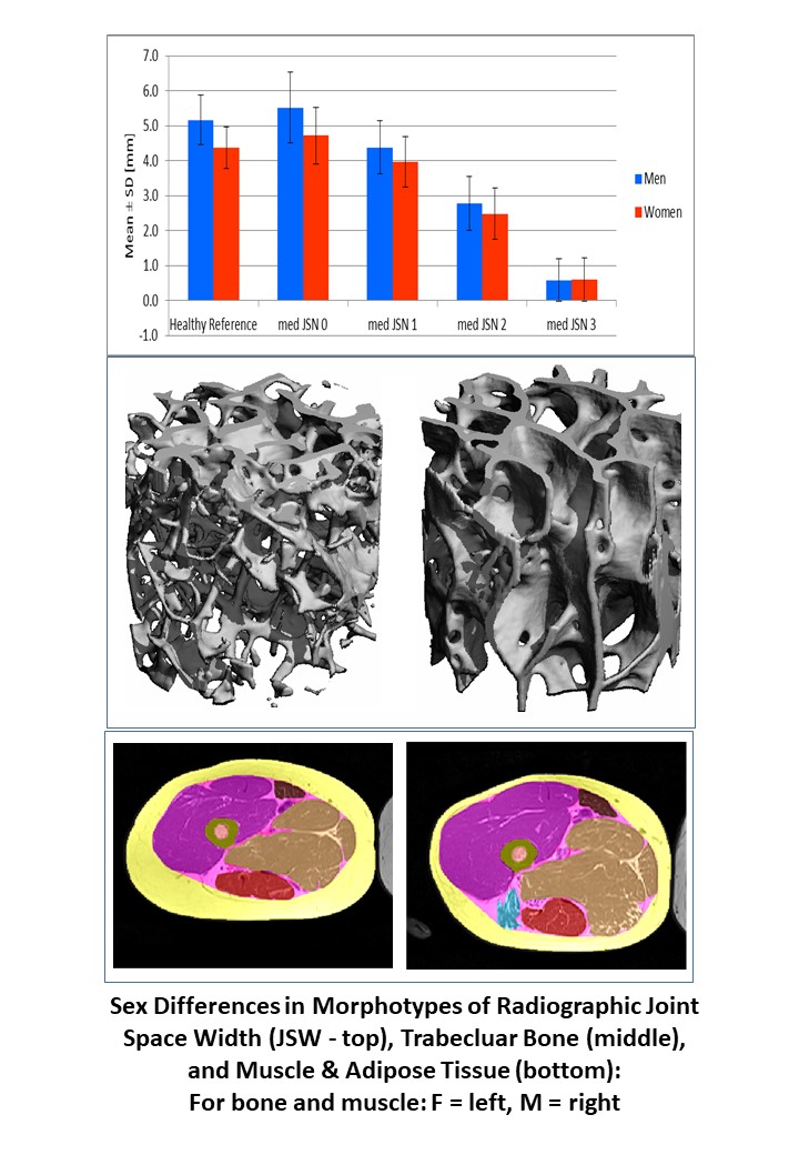
Sexual Dimorphism in Articular & Peri-Articular Tissue Anatomy – Key to Understanding Sex-Differences in Osteoarthritis? 
Osteoarthritis and Cartilage

We are excited that following a call for a Special Issue on sex differences in osteoarthritis:
https://www.sciencedirect.com/special-issue/10VMS3T8C3L with guest editors Dieuwke Schiphof (Erasmus, Rotterdam) and Tristan Maerz (University of Michigan), our two review articles on the anatomical and imaging aspects of this topic (i.e. morpho-types) are now published.

READ on sex-differences in radiographic joint space width (JSW), meniscus & ligaments, cartilage morphology, composition & deformation, and articular tissue response to treatment in the review on articular tissue anatomy in the Special Issue OAC: https://pubmed.ncbi.nlm.nih.gov/38871022/

…and then on bone size & shape, subchondral & subarticular bone, synovial membrane & infra-patellar fad-pad (IPFP), muscle & adipose tissue, and eventually peri-articular tissue response to treatment in the the second review on peri-articular tissue anatomy in OAC OPEN:  https://pubmed.ncbi.nlm.nih.gov/38946793/

  • Did you know that humans exhibit greater evolutionary homogeneity than most other species, since their genetic repertoire originates from a small population of <1500 breeding individuals, a “bottleneck” in evolution that lasted >100.000 years, bringing human ancestors close to extinction?
  • As authors, we are aware that classifications of human sex cannot be reduced to female/male. However, given lack of literature on non-binary individuals, this review article is limited to the analysis of sexual dimorphism, "morpho-types" around which variability clusters.
  • Women demonstrate more severe manifestations of musculoskeletal disorders, the inter-sex-disparity increasing with age. Knee and hand osteoarthritis are more prevalent in women, particularly symptomatic osteoarthritis.
  • It remains currently enigmatic to what extent sex-differences in osteoarthritis incidence originate from sexual dimorphism in joint anatomy, potentially exposing women to greater vulnerability.

From reviewing the literature, we conclude that female joints may be anatomically (and biomechanically) disadvantaged and at greater risk of incident knee osteoarthritis.

We reproach that only few publications mention the term “sex” in their title or abstract, data mostly “hidden” in the results.

Sex should not only be treated as a confounder, but statistical analysis should be undertaken in sex-specific strata. Sex-comparison is often confounded by demographic or disease factors.

Only few papers reported sex-differences in structure modifying interventions, this unfortunately not being the standard in clinical trials.

Sex-specific healthy reference data, and public data bases of (peri-) articular tissue properties at various developmental stages are lacking and urgently needed for comparison

The “one_size|sex fits_all” approach must be abandoned in scientific reporting. Differences between women, men, and otherwise categorized individuals must be recognized, in research as well as in clinical practice.

This approach should be pursued per self-commitment of the scientific-community, and adopted into official editorial policies of scientific journals and text books for full implementation.

We are tremendously grateful to the many colleagues who supported us in identifying and reporting these data:

Article 1 (OAC Special Issue): Gerard Ateshian, Francis Berenbaum, Flavia Cicuttini, Martin Englund, Ali Guermazi, David Hunter, Mylène Jansen, Gabby Joseph, Thomas Link, Malcolm Logan, Michael Nevitt, Frank Roemer

Article 2 (OAC OPEN): Kim Bennell, Francis Berenbaum, Mikael Boesen, Alan Brett, Ali Guermazi, David Hunter, Gabby Joseph, Olga Kubassova, Virginia Kraus, Thomas Link, Steve Messier, Ralph Mueller, Michael Nevitt, Andrew Pitsillides, Frank Roemer, Kathryn Stok


SUMMARY & POSTERS
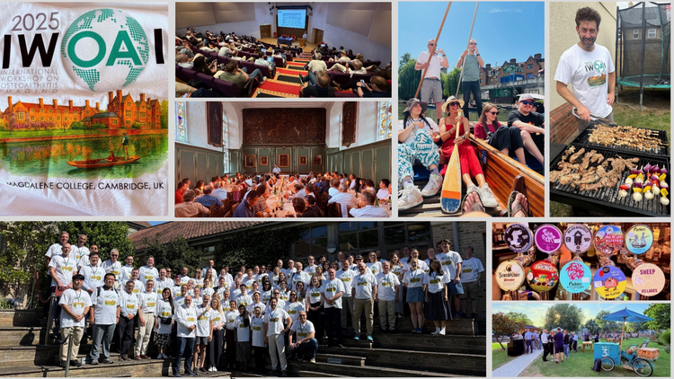
INTERNATIONAL WORKSHOP ON OSTEOARTHRITIS IMAGING (IWOAI), JUNE 25-28, 2024

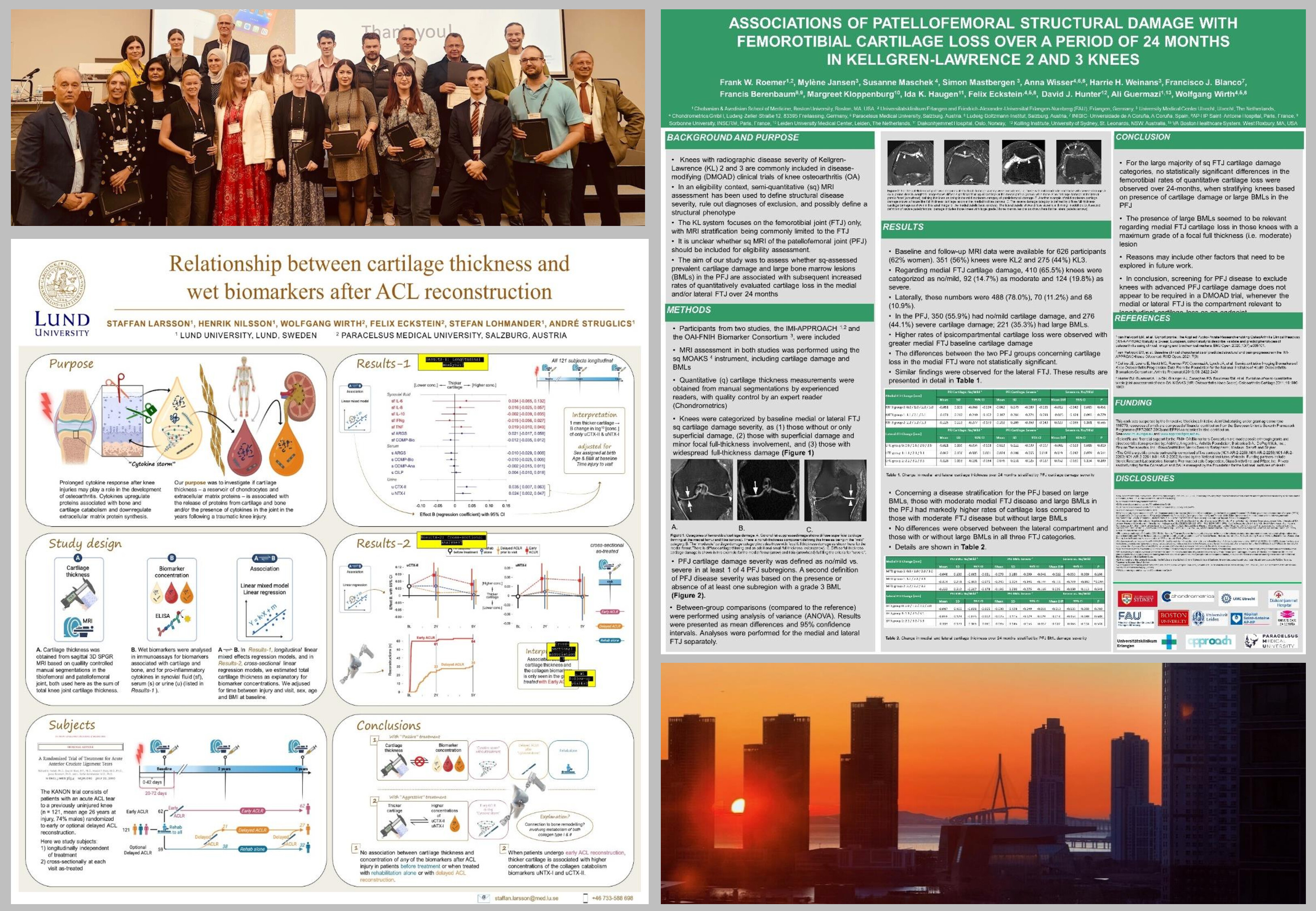
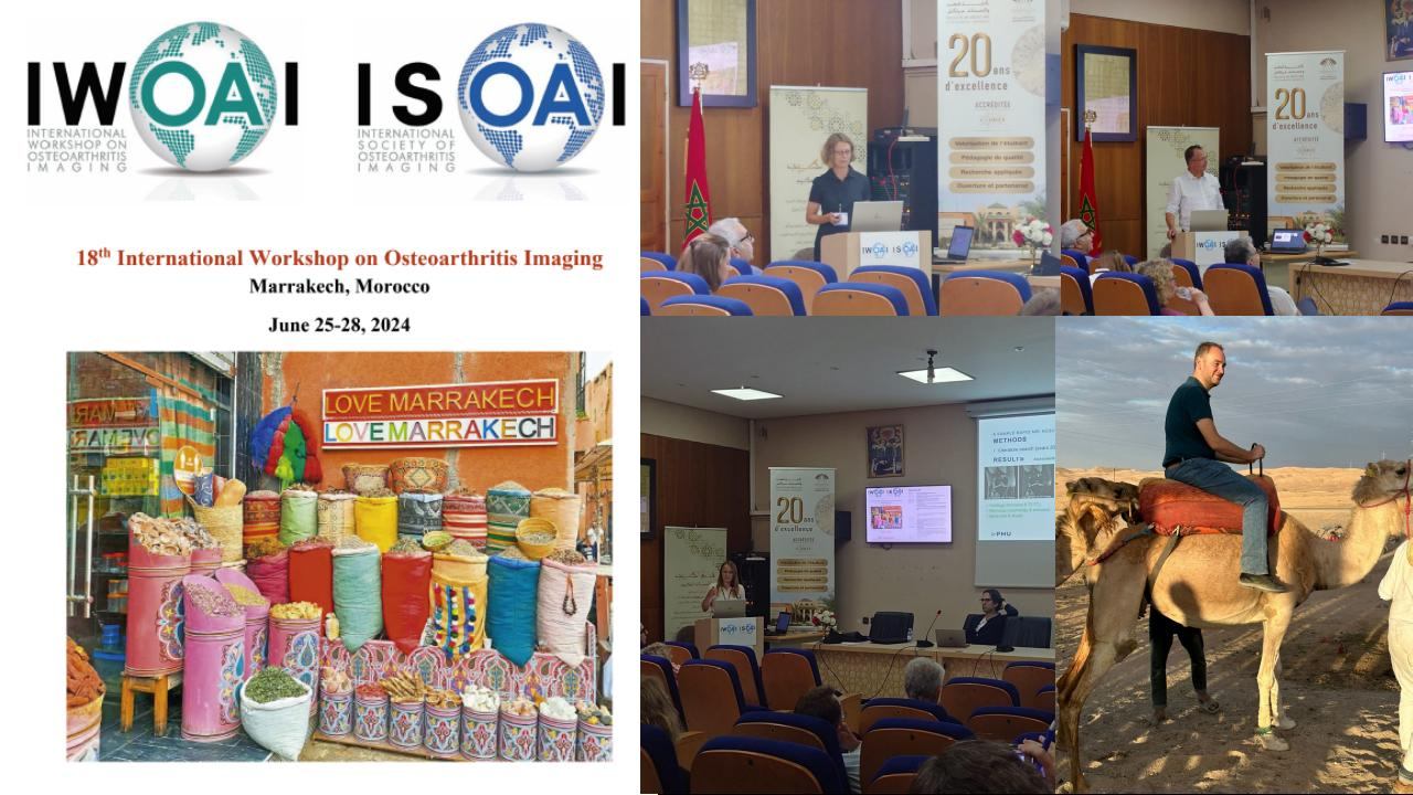
The team of Chondrometrics GmbH and the Research Program for Imaging at Paracelsus Medizinische Privatuniversität presenting at the IWOAI in Marrakech, Morocco

We spent 4 rich and intense days at the 2024 International Workshop of Osteoarthritis Imaging (18th IWOAI) in Marrakech, Morocco, with enlightening lectures on the relationship between pain and structure in osteoarthritis. There was great opportunity to establish new contacts and nurturing existing friendship amongst researchers from academia and industry, hopefully soon bringing the first and long-awaited disease modifying osteoarthritis drug (DMOAD) to market.

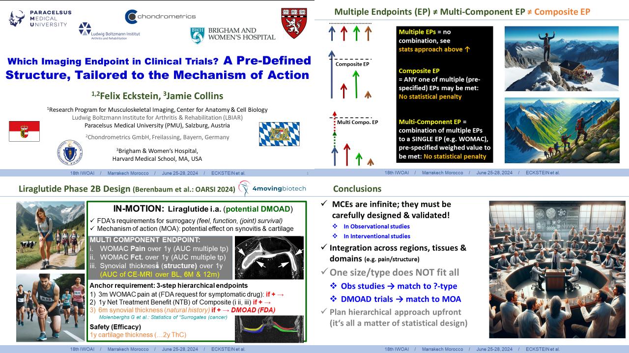
Felix Eckstein with Jamie Collins were invited to speak about multi-component and composite outcomes vs. single endpoints tailored to the specific mechanism of action (MOA) in putative DMOAD trials. This will be followed by a “Perspective” on this topic in the “Osteoarthritis Imaging” Journal (OSTIMA) and will be hopefully available for reading soon.

IWOAI Marrakesh Eckstein

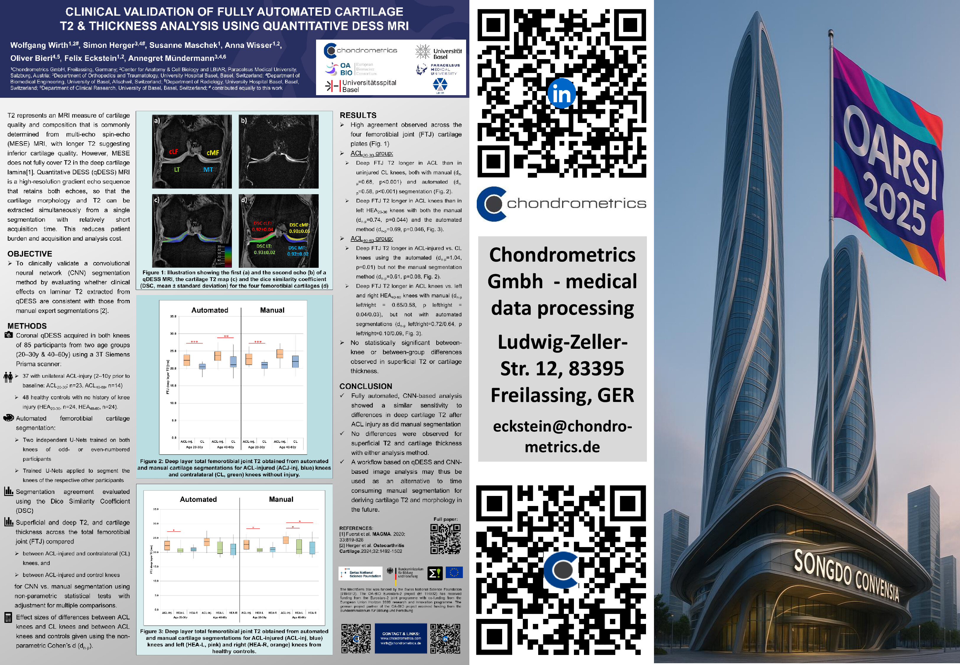
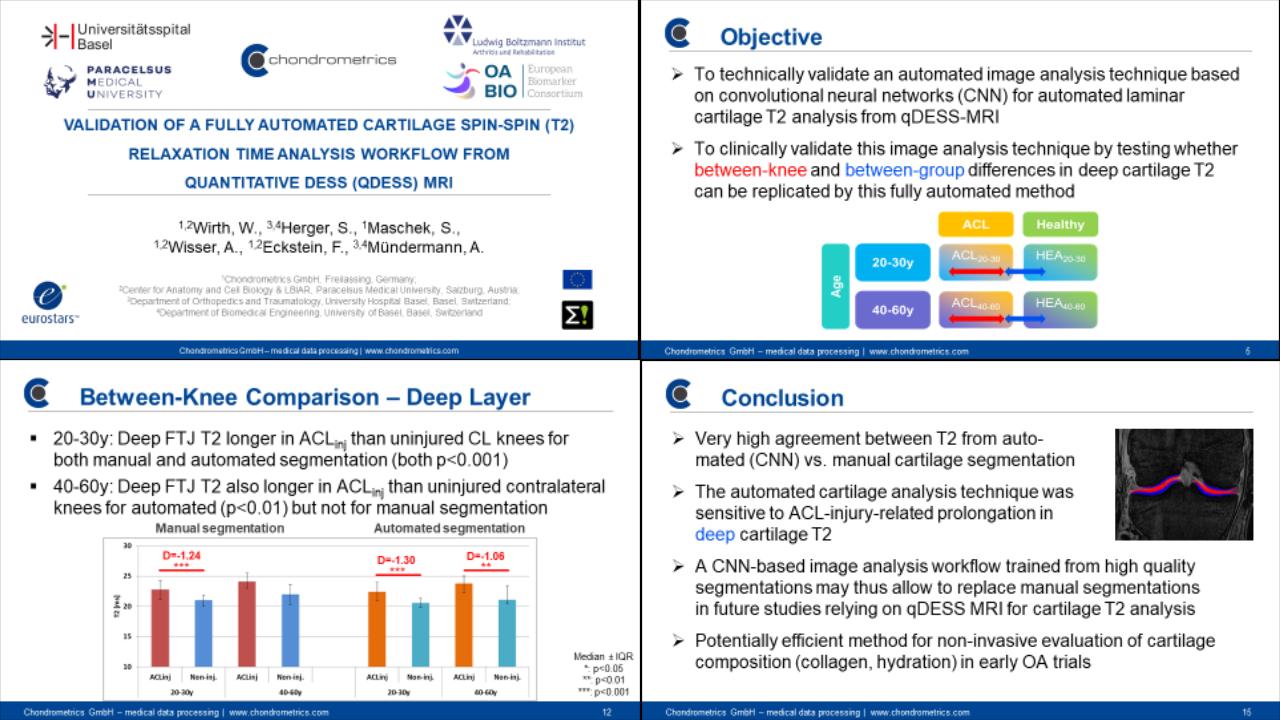
Wolfgang Wirth presented on the clinical validation of cartilage transverse relaxation time by MRI (T2), known to be related to cartilage composition and mechanical properties. Segmentation was performed using new fully automated convolutional-neural-network (CNN)-based models, and a novel high-resolution qDESS MRI sequence. The study was conducted in close collaboration with Annegret Mündermann and Simon Herger from the University of Basel, Switzerland. It was found that knees with anterior cruciate ligament (ACL) injury exhibit longer T2 (suggesting early cartilage pathology) in the deep femorotibial cartilage compared with contra-lateral knees or healthy reference knees. The superficial cartilage T2 and the cartilage thickness, in contrast, were not significantly different between case and reference knees. This work was supported by a Eurostars OA-BIO consortium grant and represents a big step forward in achieving time- and cost-efficient analysis of cartilage T2 and thickness from the same image acquisition (qDESS) and (automated) segmentation algorithm.

IWOAI Marrakesh Wirth

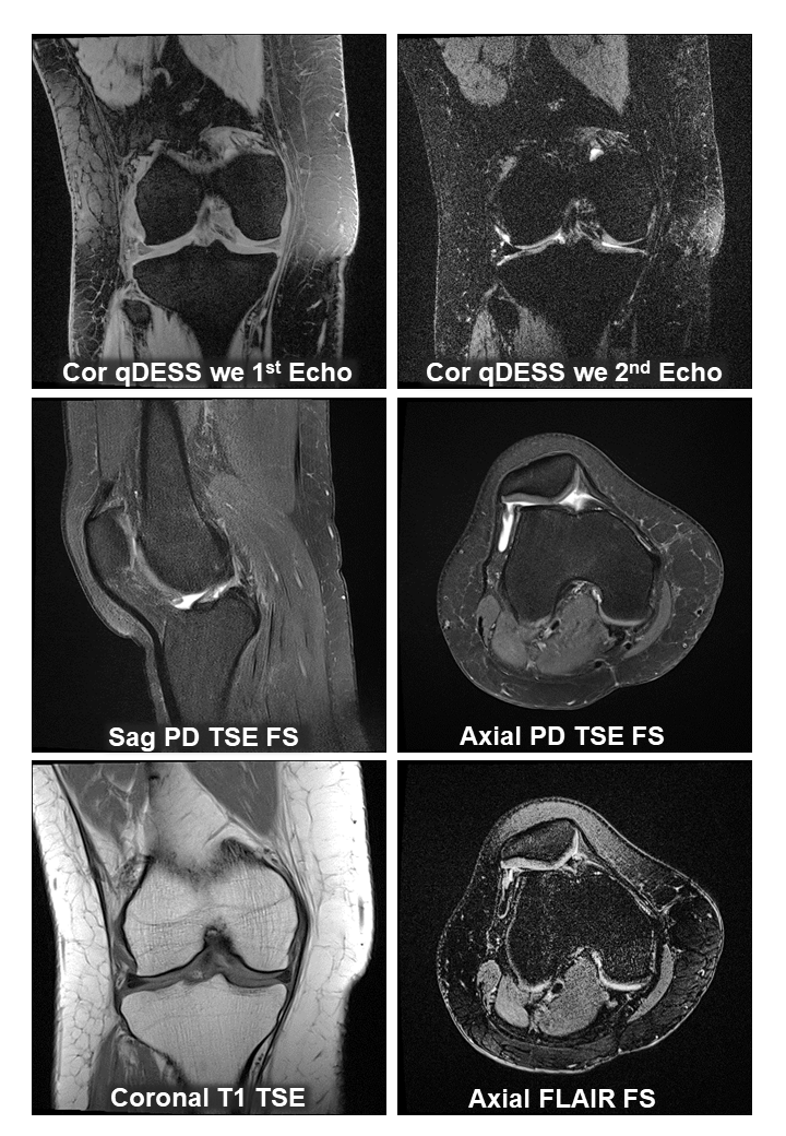
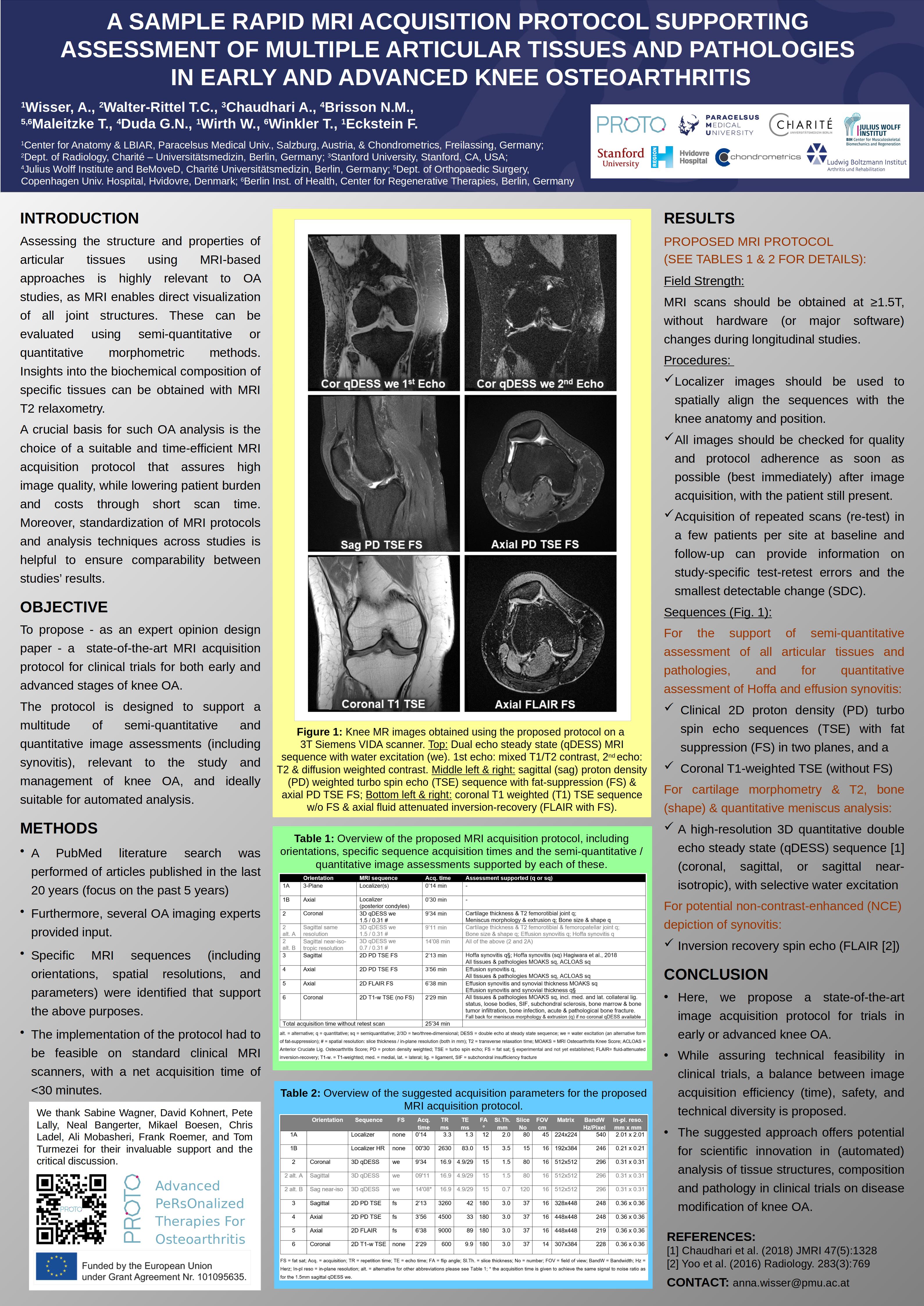
Anna Wisser displayed a poster on a rapid (<30 min) sample acquisition protocol, encompassing MRI sequences for (quantitatively) assessing synovitis (no contrast), cartilage thickness and cartilage T2, and (semi-quantitative scored) structural pathologies of articular tissue. This first study in the PROTO Horizon project was co-authored by Chondrometrics GmbH team members and Nicholas Brisson, Georg Duda, Tazio Maleike, and project head Tobias Winkler from the Universitätsmedizin Berlin.

IWOAI Marrakesh Wisser

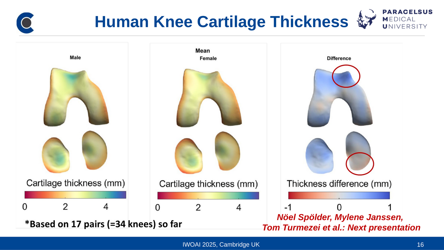
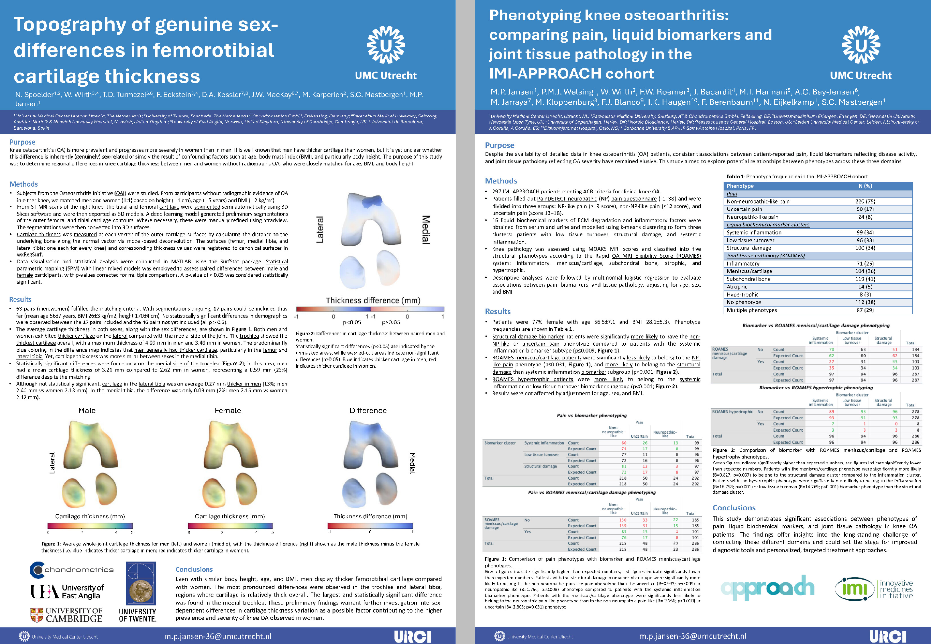
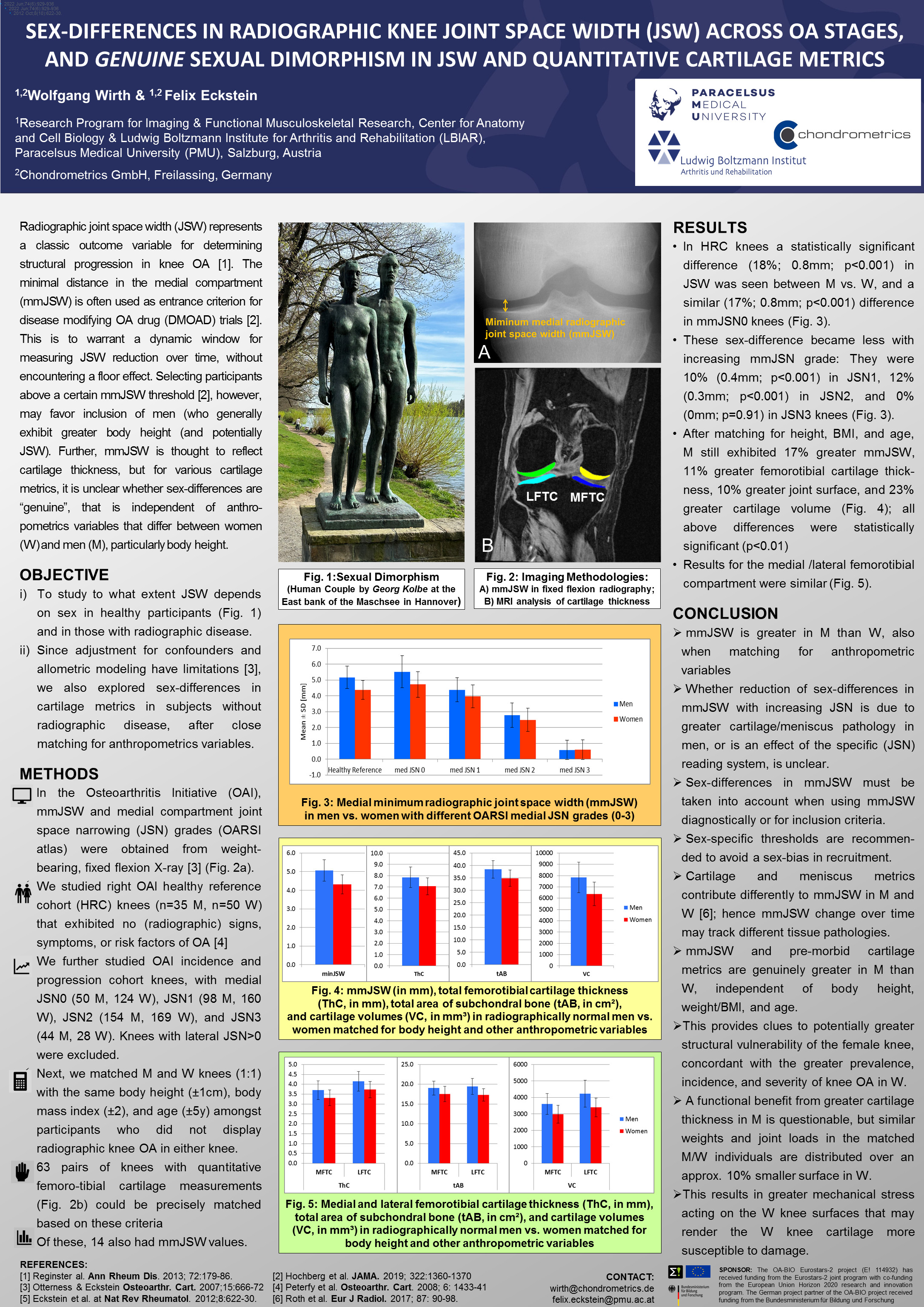
Susanne Maschek presented a poster on sexual dimorphism of radiographic joint space width (JSW) in joint space narrowing (JSN) strata of almost 1000 subjects. Med higher values of greater JSW than women, but the difference decreased with greater JSN. She further assessed "genuine" sex differences in femorotibial cartilage thickness, independent of other demographic factors. Women matched for the same body height-, age-, and BMI and without radiographic disease values than women still displayed approx. 10% less cartilage thickness and also smaller joint surface areas, indicating that women may be structurally disadvantaged in terms of suffering from knee osteoarthritis (for the poster, please see the OARSI 2024 NEWs section)

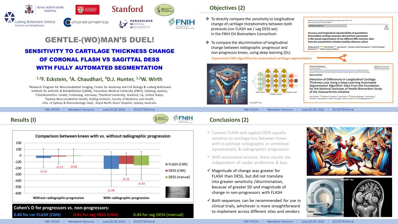
Felix Eckstein eventually compared the performance of coronal FLASH and sagittal DESS MRI in discriminating progressor vs. non-progressor knees in the OAI FNIH Biomarker sample (FNIH1), using fully automated segmentation of cartilage thickness. Co-authored by Wolfgang Wirth from Chondrometrics, Akshay Chaudhari (Standford University) and David Hunter (University of Sydney), the novely of that study is that the comparison is without potential reader preference or bias, given that the analysis was done fully automated using a CNN-based deep learning AI algorithm that was trained on the FLASH and DESS images of a different set of knees. The study reported greater rates of progression by FLASH than DESS, but also greater standard deviations and progression in control subjects, so that the actual discrimination of both groups was very similar across both MRI sequences

IWOAI Marrakesh Eckstein Oral

Impressions from Marrakesh

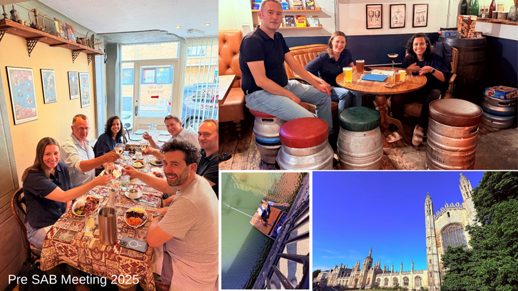
Ali Guermazi, MD, PhD Guermazi is to be hugely thanked for organizing a fantastic conference and venue that kept us without sleep not only for the day but also for most of the nights. We all look forward to meet in Cambridge for the 19th IWOAI hosted by Tom Turmezei in July 2025.

IWOAI Marrakesh Impressions IIWOAI Marrakesh Impressions II


EVENT & POSTERS
EULAR CONFERENCE VIENNA, JUNE 12-15, 2024

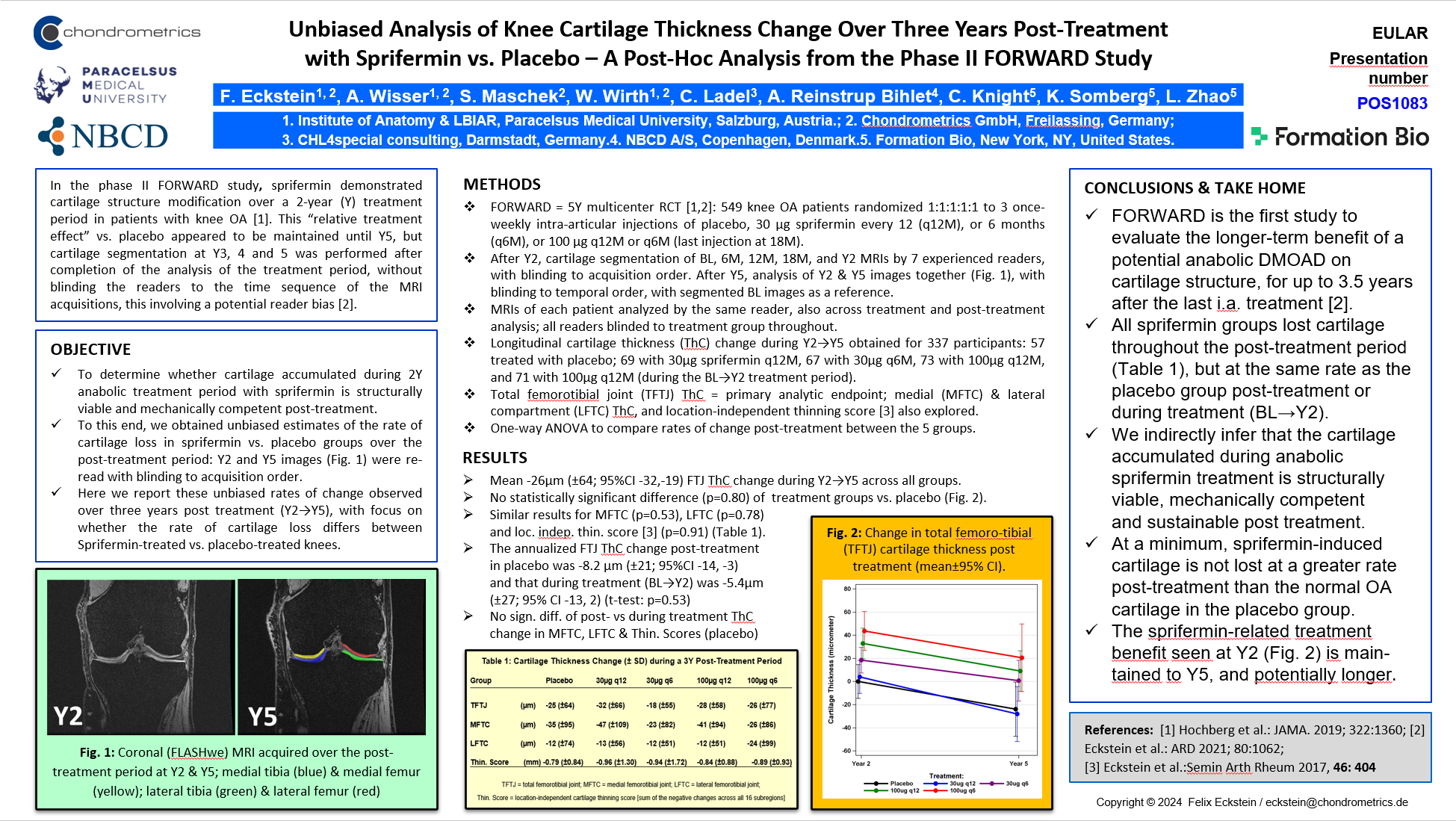
The team of Chondrometrics GmbH and partners present a poster titled: "Unbiased Analysis of Knee Cartilage Thickness Change Over Three Years Post-Treatment with Sprifermin vs. Placebo – A Post-Hoc Analysis from the Phase II FORWARD Study"
EULAR Announcement

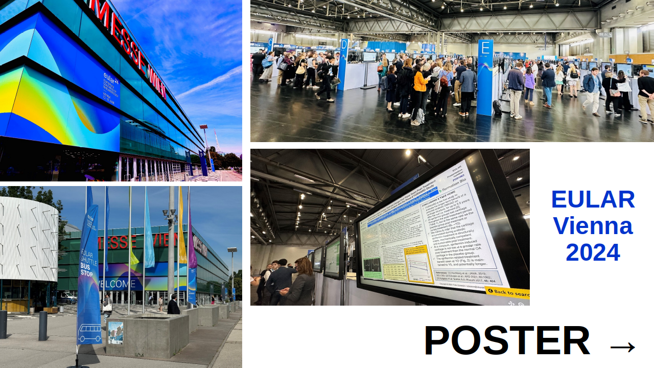
The “European Alliance of Associations for Rheumatology” (EULAR) is one of the largest organizations of its kind worldwide, and holds one of the largest research conferences in rheumatoid and osteoarthritis. This year, the conference takes place from June 12-15 in beautiful Vienna, with more than 18,000 participants from over 130 countries expected to attend EULAR Congress.

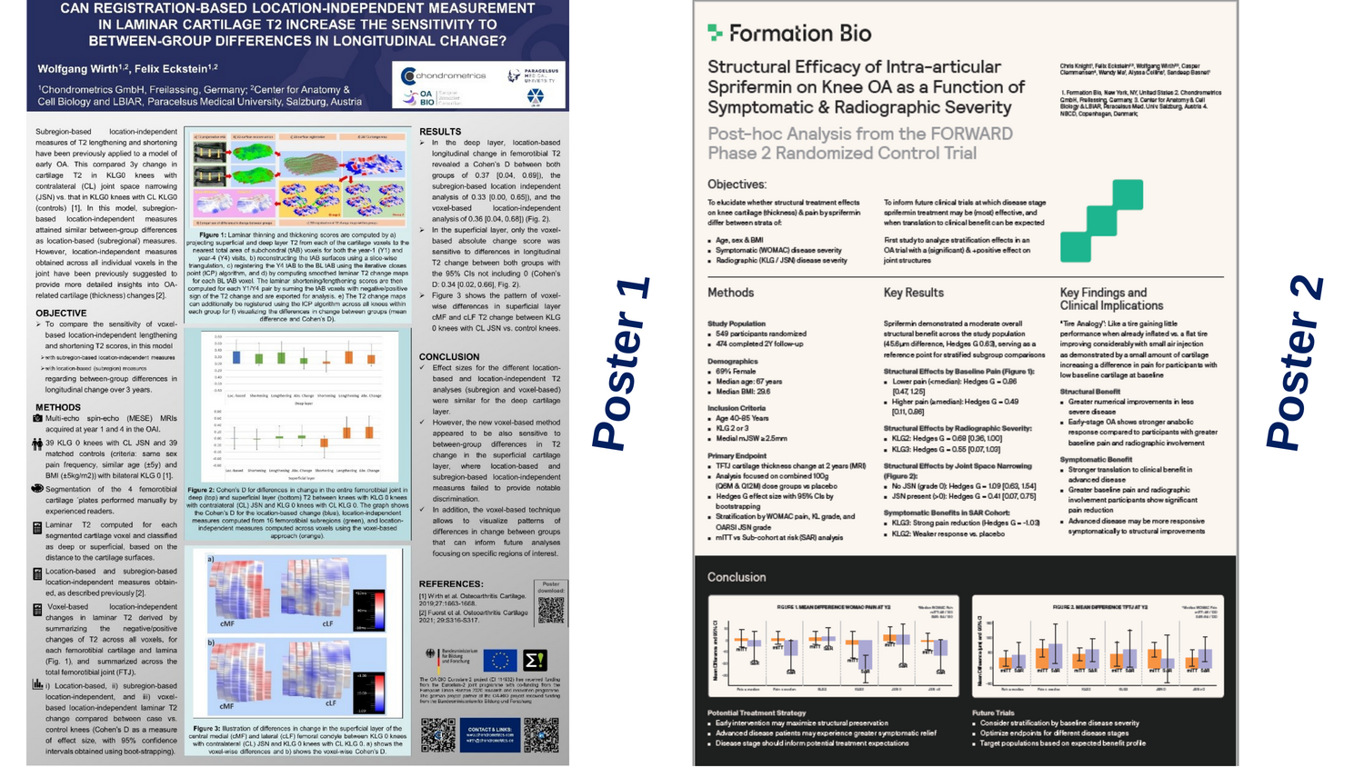
Two weeks from now (Friday, June 14, at 12:00 CEST), we will - with our esteemed colleagues from Formation Bio, NBCD, and CHL4special consulting - present a poster titled: “Unbiased Analysis of Knee Cartilage Thickness Change Over Three Years Post-Treatment with Sprifermin vs. Placebo – A Post-Hoc Analysis from the Phase II FORWARD Study”. #POS1083 will be displayed during the Poster Tour: “Clinical Studies in Osteoarthritis” (Room Poster View / Session VI).

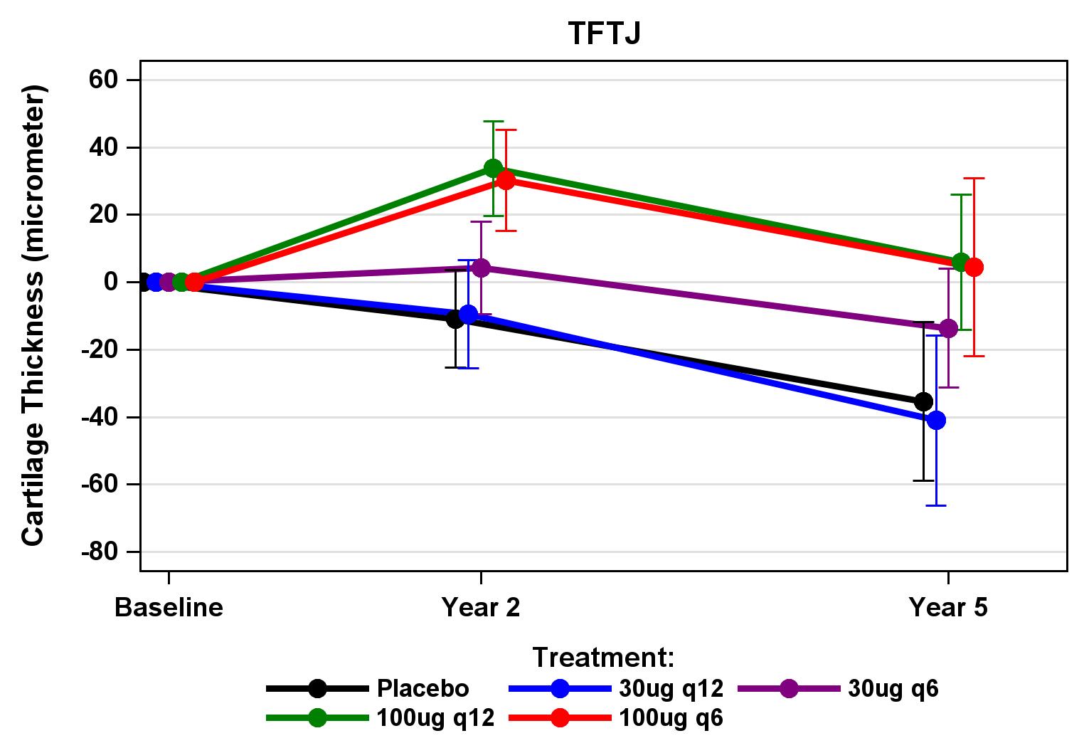
Cartilage Thickness TFTJ

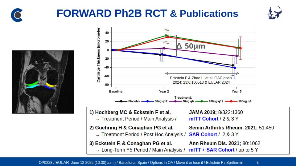
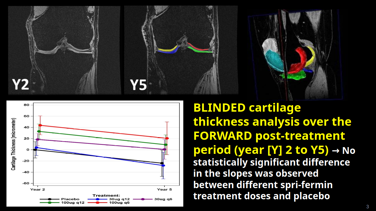
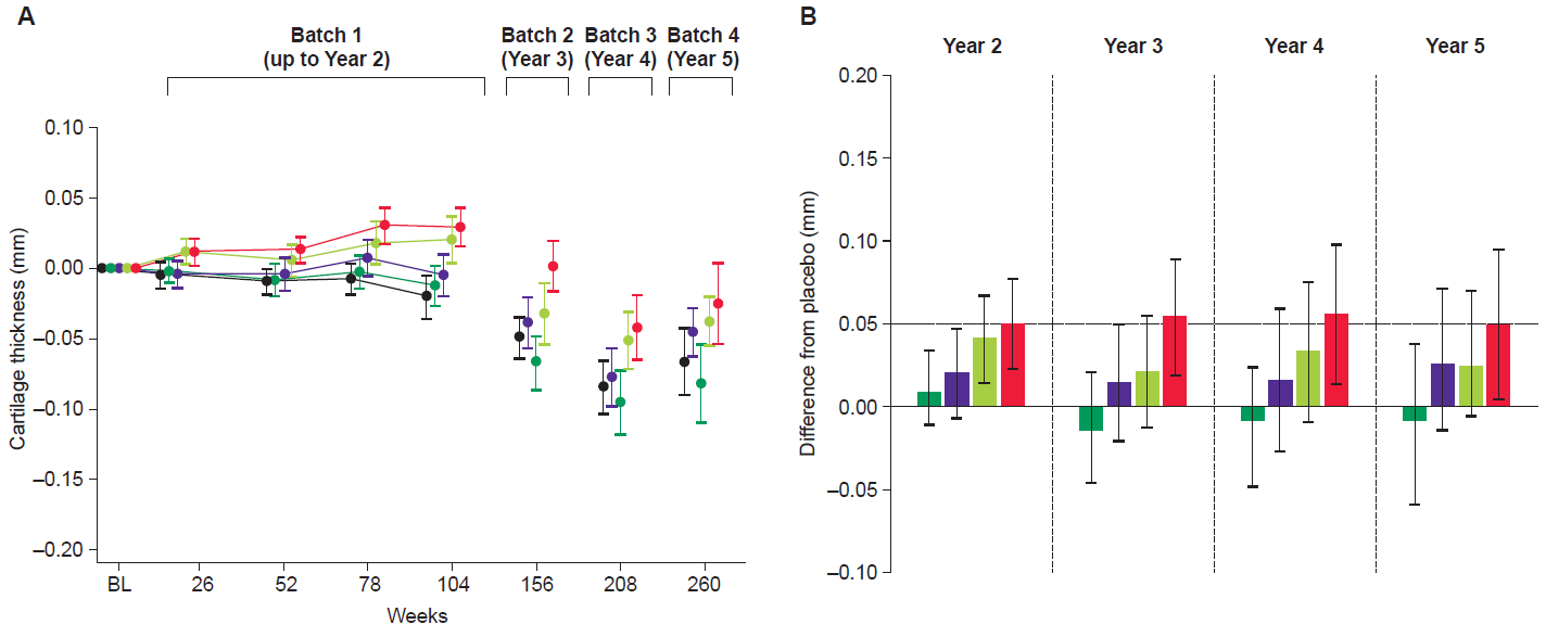
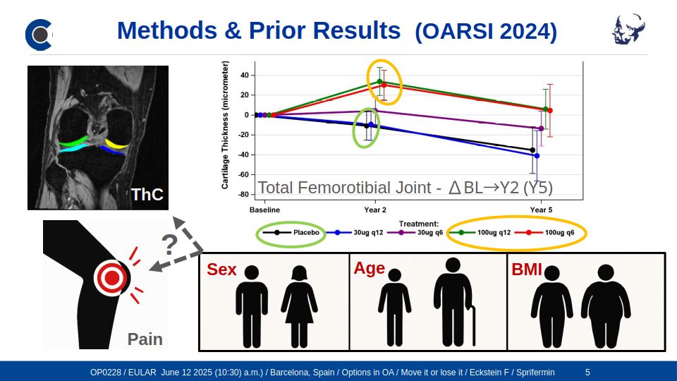
Figure showing the quantitative change in cartilage thickness by MRI morphometry in the (total) femorotibial joint (TFTJ) during the treatment phase (baseline to Y2), and during the post-treatment period (Y2 to Y5). The readers were blinded to the relative temporal order of the MRIs for both periods: q12 = drug injected i.a. every 12 months, and q6 = every 6 months (up to 18 months), at dosages of 30 µg or 100 µg, respectively.

The FORWARD study (NCT01919164) is one of the first placebo controlled, randomized control trials (RCTs) that have clearly demonstrates an anabolic effect of a drug (Sprifermin) on joint structure in knee osteoarthritis, specifically cartilage thickness by magnetic resonance imaging (MRI). And the first to include an extensive follow-up period of 3 years (Y) after a 2Y treatment period (Fig.). For a summary and “download area” of original publications related to Sprifermin, please visit the research section of our Webpage: Sprifermin Summaries > Exclusive Executive Summaries > The Sprifermin Program.

At EULAR 2024, we will present an unbiased analysis of the FORWARD post-treatment MRI data on articular cartilage thickness change, measured using quantitative morphometry by Chondrometrics 3.0 software. Expert readers were blinded to the temporal order of the MRI acquisitions acquired at Y2 and Y5. Our results demonstrate that the rates of cartilage thickness loss (slopes) during this period do not differ significantly between placebo and the various sprifermin dose groups (Fig.). Further, the difference in cartilage thickness seen at Y2 between sprifermin-treated participants and placebo is fully maintained up to Y5 (Fig.). The additional sprifermin-induced cartilage tissue present at the end of the treatment period is hence not lost at greater rates post-treatment than “normal” osteoarthritic cartilage of placebo participants. This supports the interpretation that cartilage tissue gained during anabolic treatment is biologically viable and mechanically competent post-treatment, warranting sustainable structural treatment effects on knee osteoarthritis for several years.

Join us on Friday, June 14, 2024, noon, for more data and lively discussions.

Cartilage Thickness TFTJCartilage Thickness TFTJCartilage Thickness TFTJCartilage Thickness TFTJ


SUMMARY & POSTERS
OARSI WORLD CONGRESS VIENNA, APRIL 18-21, 2024

The team of Chondrometrics GmbH and the Research Program for Imaging at Paracelsus Medizinische Privatuniversität presenting at the World Congress in Vienna

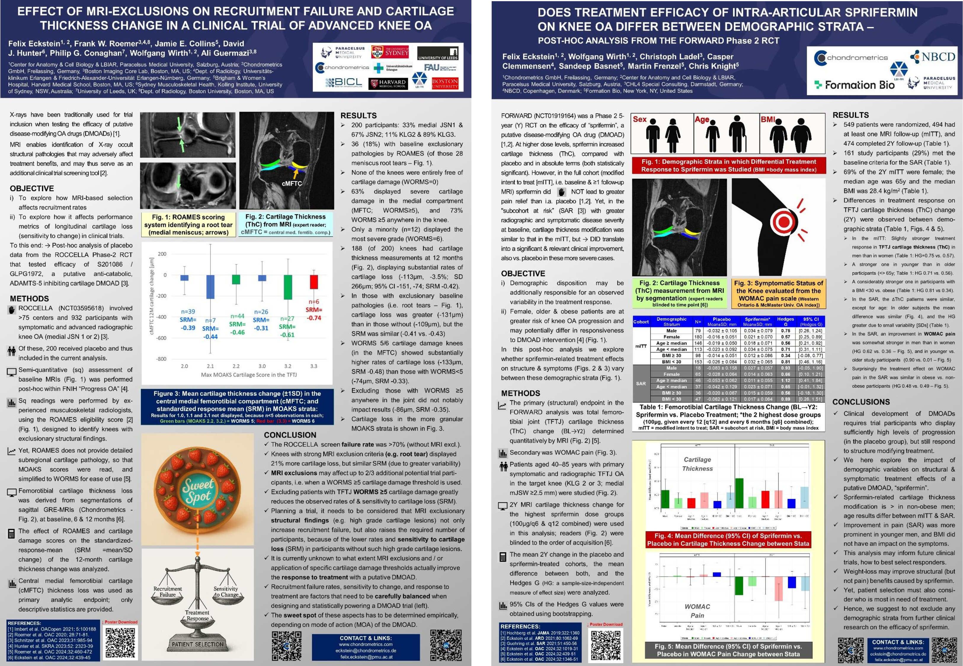
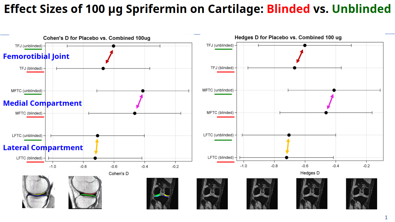
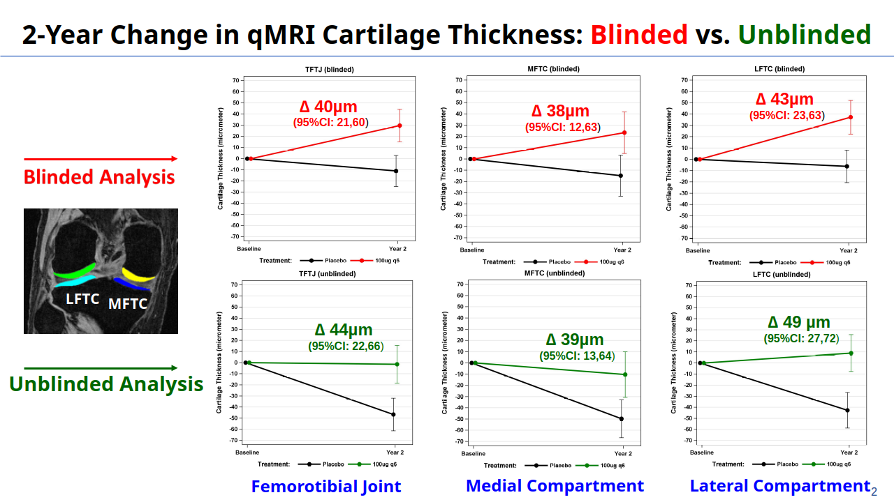
The team of Chondrometrics GmbH and the Research Program for Imaging at Paracelsus Medizinische Privatuniversität presenting at the World Congress in Vienna included Susanne Maschek (Chief of Human Resources and Quality Control), Anna Wisser (Project Manager), Wolfgang Wirth (Chief Technical Officer and Chief of Quality Control), Clemens Heistracher (PhD Student), and Felix Eckstein (CEO & CMO). Felix Eckstein gave an Oral Presentation during the Imaging Session on Friday, April 19th (No. 20) on the impact of unblinded reading vs. segmentations blinded to the order (time point) of image acquisition on the rates of longitudinal cartilage thickness change, and on the structural treatment effects on cartilage in osteoarthritis.
During the China-Germany-Night later the same day, he gave a Review Lecture on the role of imaging biomarkers in the development of disease modifying osteoarthritis drugs (DMOADs), on current disease-modifying treatment trials in osteoarthritis, and on recent results obtained with fully automated segmentation & analysis of articular tissues, using AI- (more specifically CNN-) based algorithms.
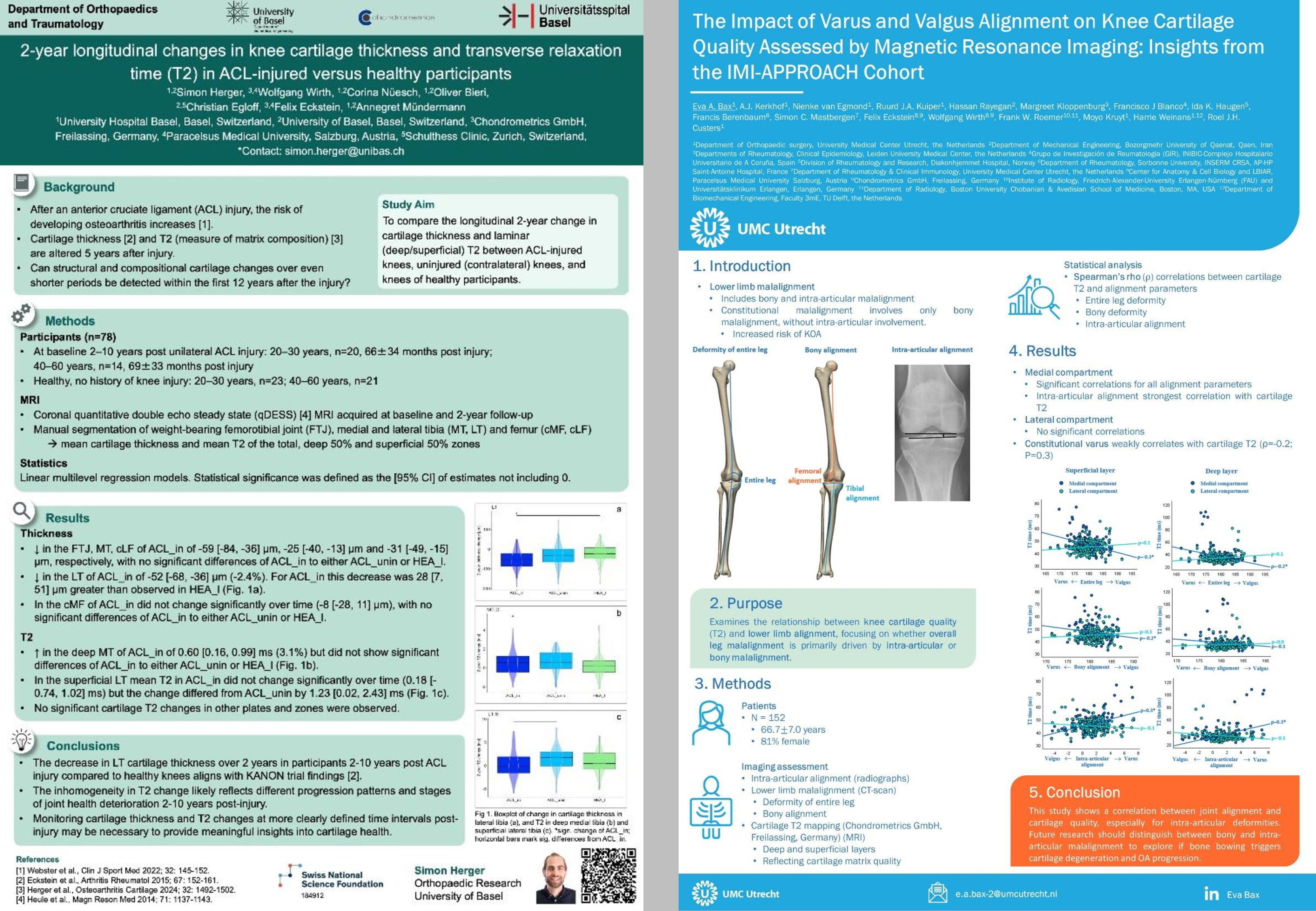
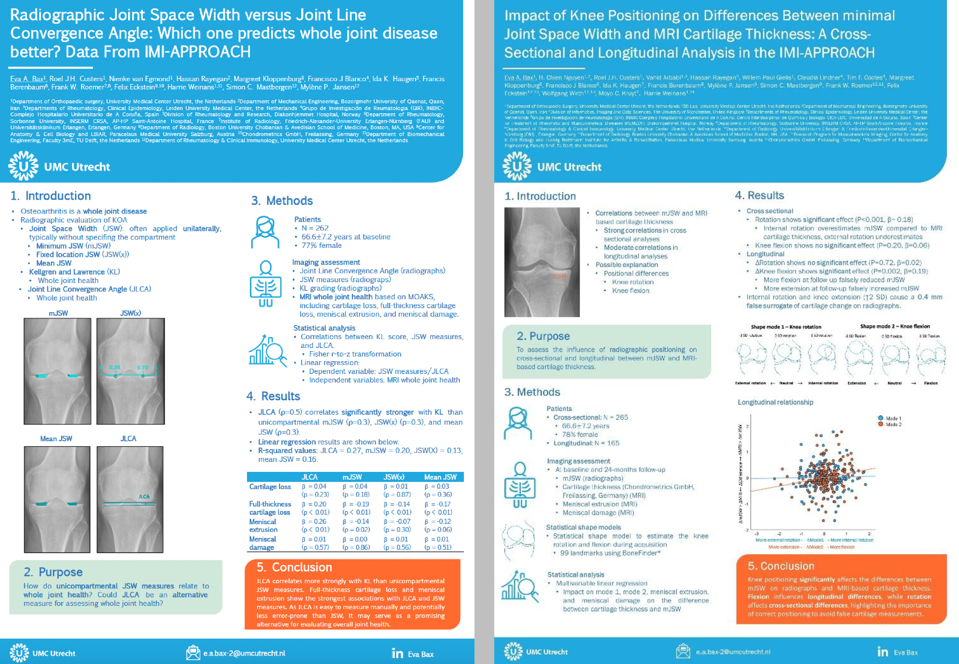
The group presented 6 first-author posters, and co-authored another 4 posters with their collaborating partners, all displayed below. The 10 posters cover a wide range of imaging applications in osteoarthritis research and nicely reflect the current status of work both at Chondrometrics and at the group at PMU.
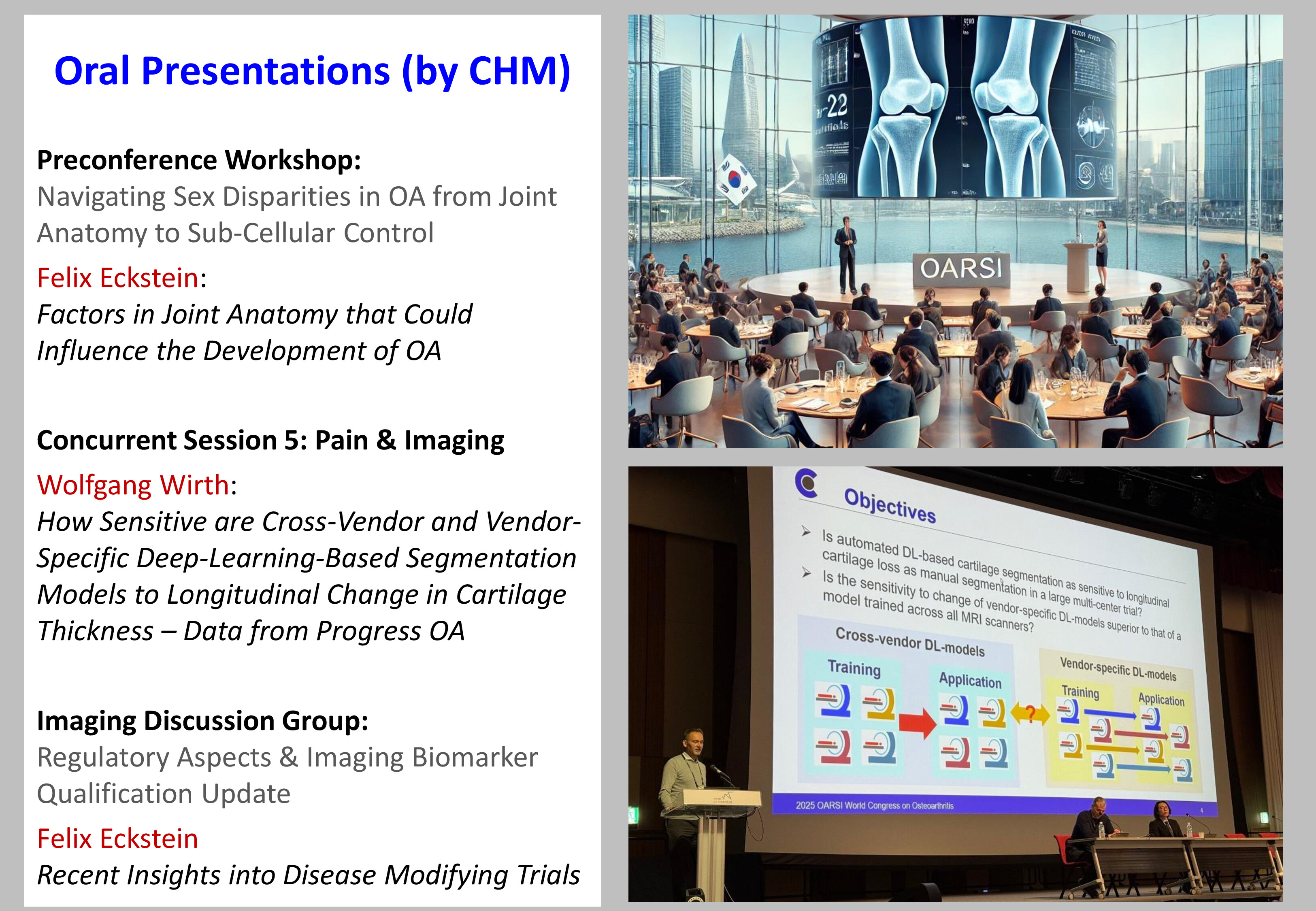
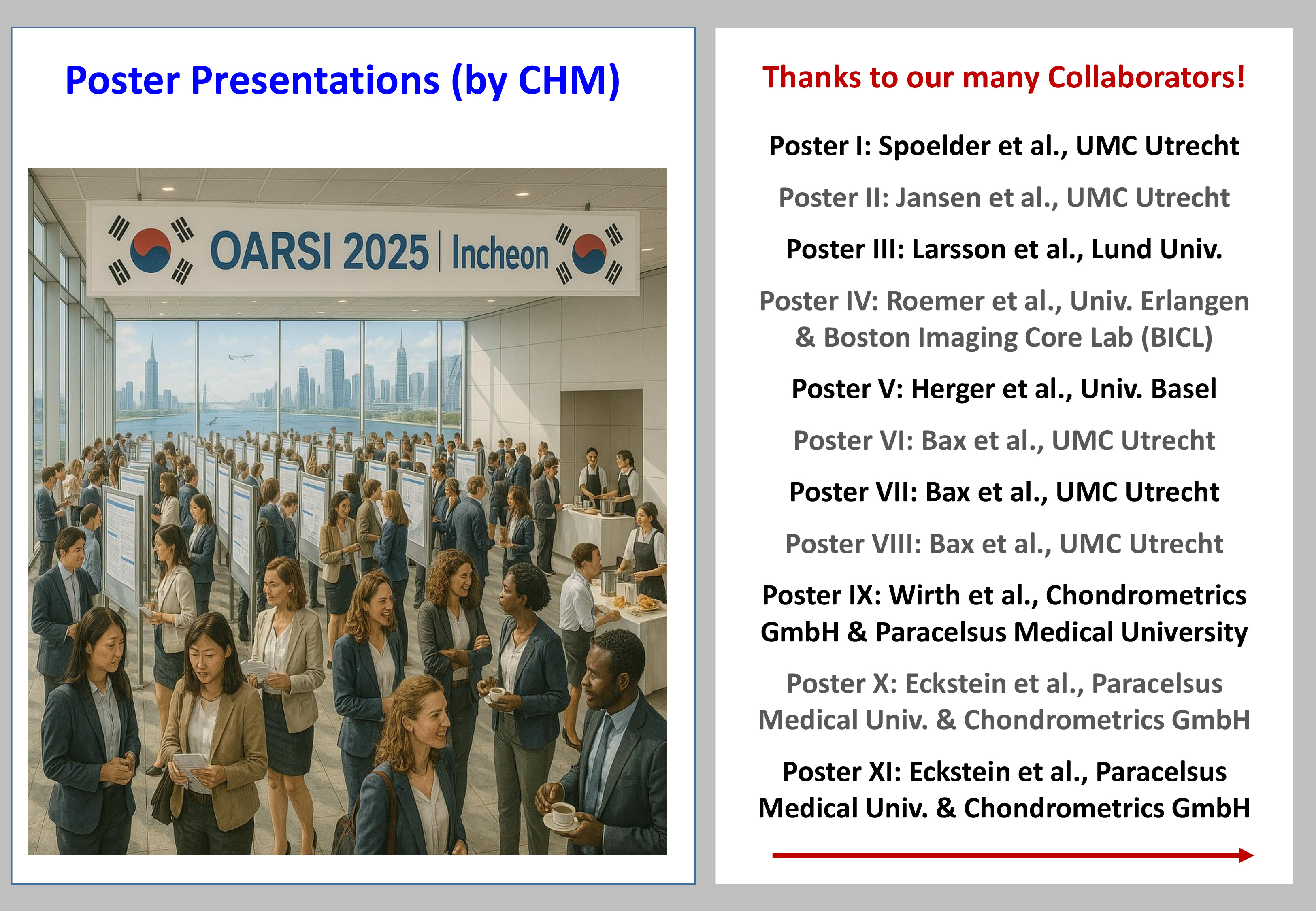
We hope seeing you again at OARSI 2025 next April in Seoul, Korea.
Finally we would like to thank the below researchers for their invaluable contributions and the exciting collaborations:


First-author posters

Thanks to:   Frank Roemer, Simon Lévy, Armin Nagel, Maximilian Bachl, Michael Uder

Thanks to:  Akshay Chaudhari, David Hunter

Thanks to: Nicholas Brisson, Georg Duda, Francis Berenbaum

Thanks to: Christina Niedling, Pætur Holm, Søren T. Skou, Jana Eder (Kemnitz)


Co-author posters

Thanks to: Jamie Collins, Elena Losina, Thomas Fuerst, Douglas Robinson, Ali Guermazi, Peter Mesenbrink, Letizia Deveza, David Hunter, Virginia Kraus, Christoph Ladel, Thomas Perry, Christopher Swearingen, Erik Dam

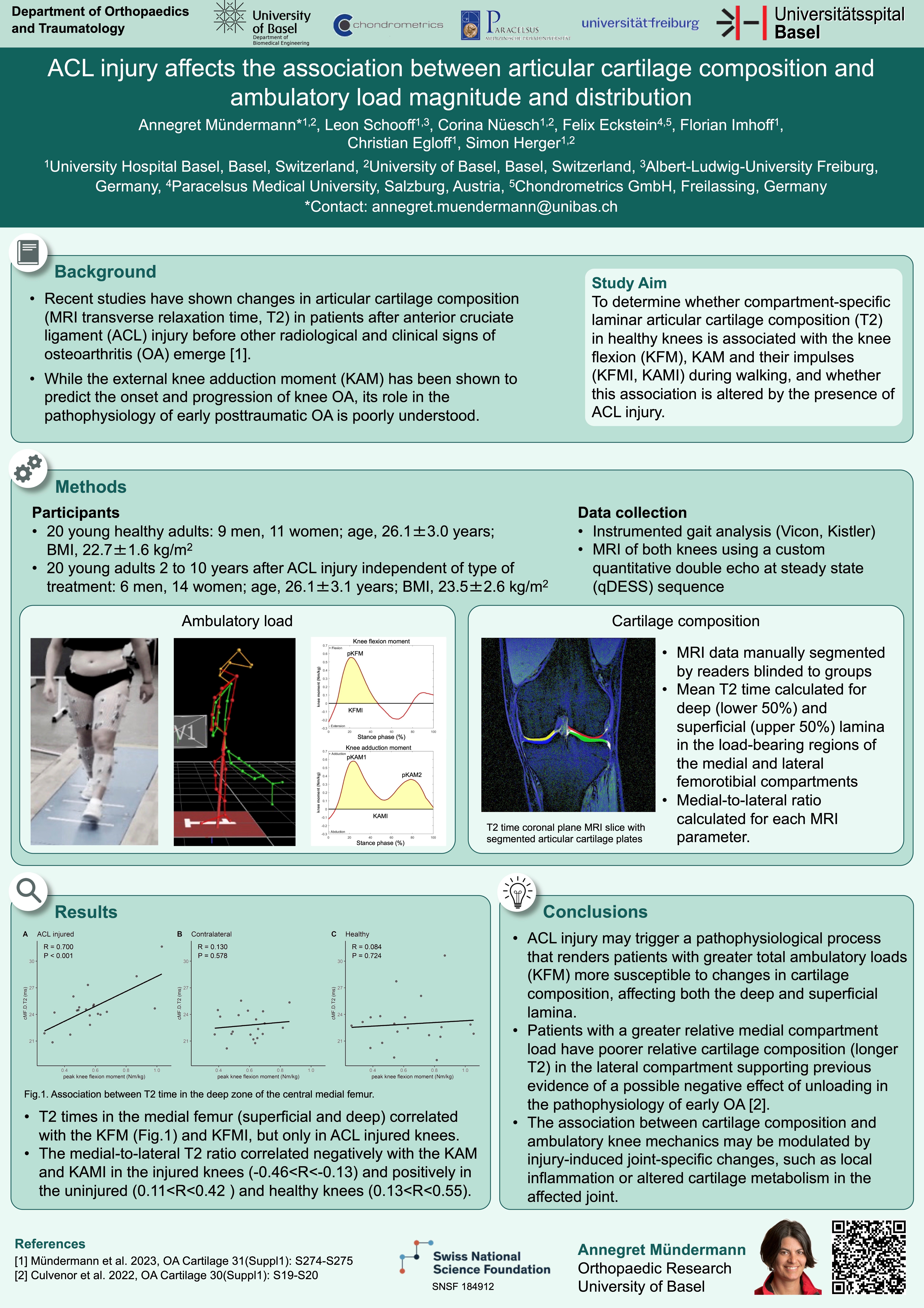
Thanks to: Annegret Mündermann, Leon Schoof, Corina Nüesch, Florian Imhoff, Christian Egloff, Simon Herger

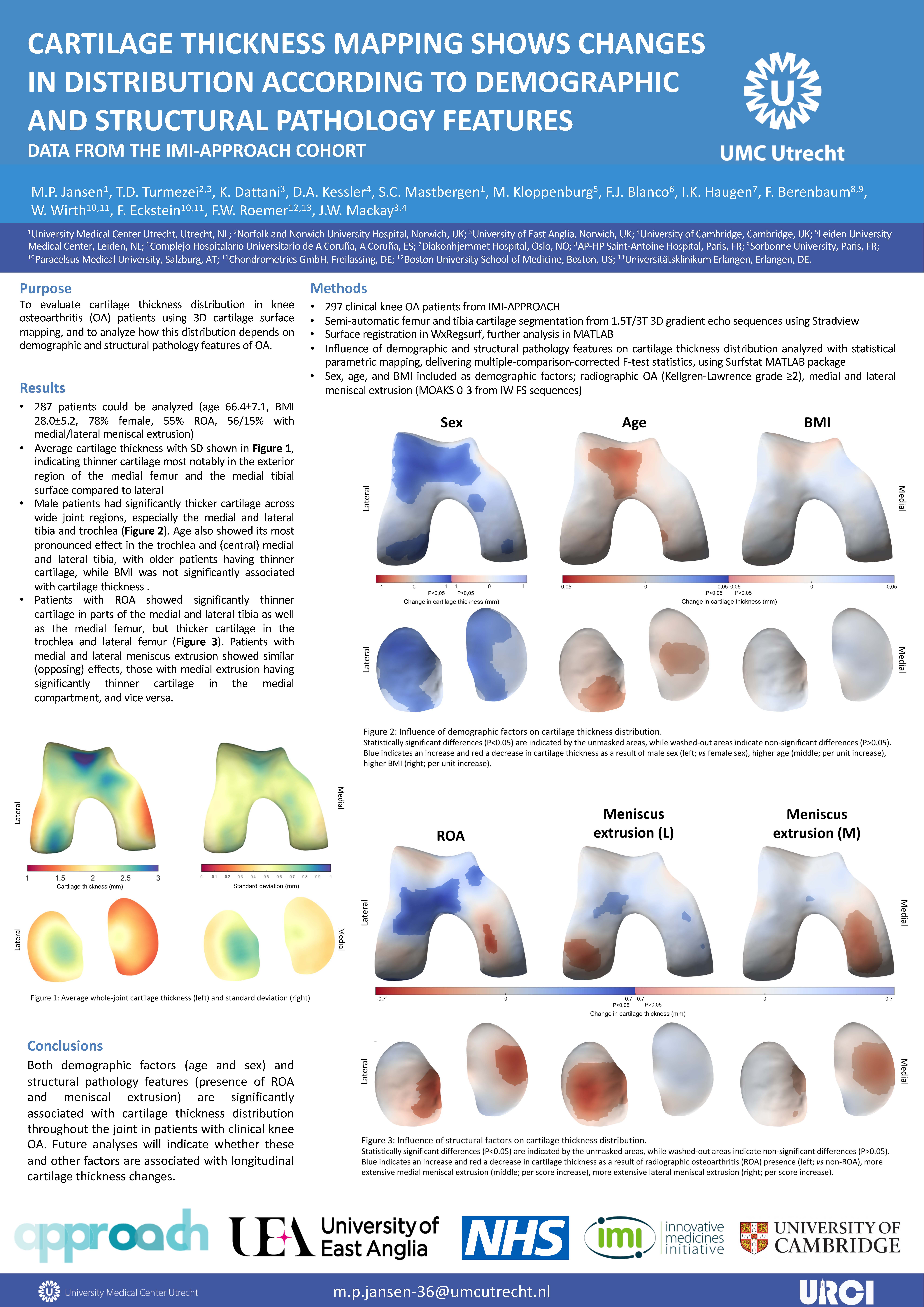
Thanks to: Mylène Jansen, Simon Mastbergen, Tom Turmezei, Kishan Dattani, Jamie MacKay, Margreet Kloppenburg, Dimitri Kessler, Francisco Blanco, Ida Haugen, Francis Berenbaum, Frank Roemer 

Thanks to:  Eva Bax, Chien Nguyen, Roel Custers, Nienke van Egmond, Vahid Arbabi, Hassan Rayegan, Willem Paul Gielis, Claudia Lindner, Tim F. Cootes, Margreet Kloppenburg, Francisco J. Blanco, Ida K. Haugen, Francis Berenbaum, Mylène P. Jansen, Simon C. Mastbergen, Harrie Weinans


ROAD TRIP
TAKING THE LONG WAY TO OARSI, APRIL 12-15, 2024

David Hunter and Felix Eckstein to Vienna via 4 rivers and by bike 

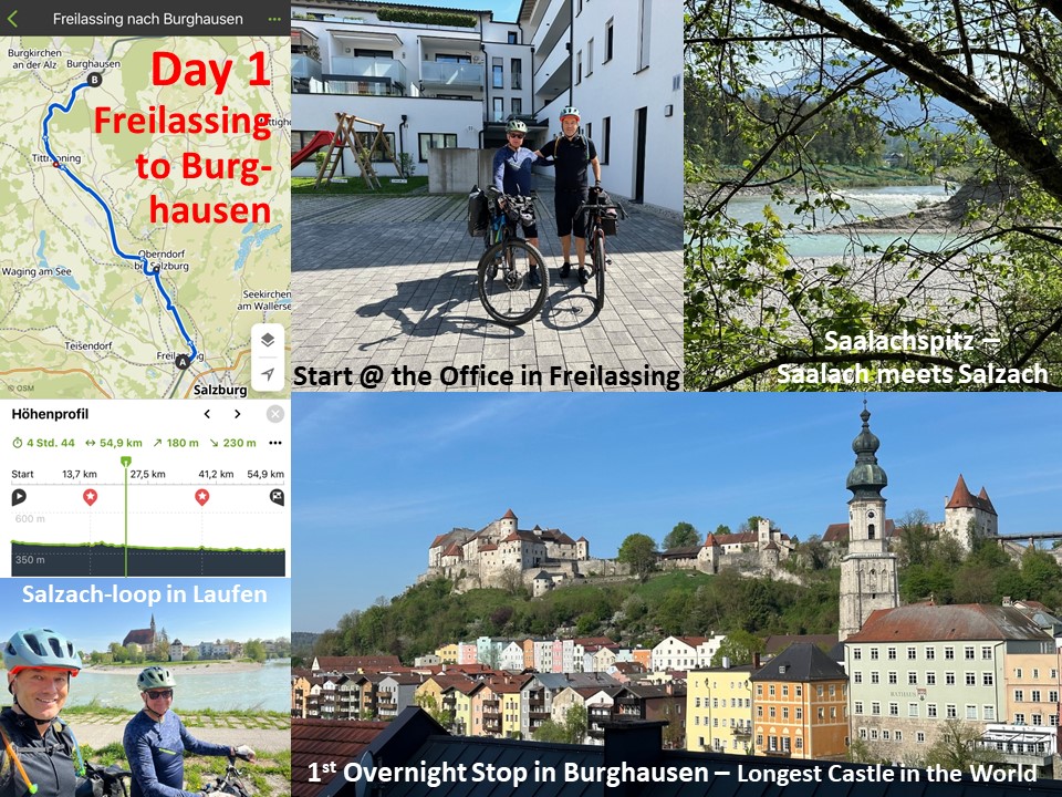
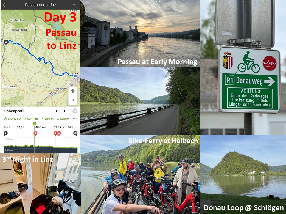
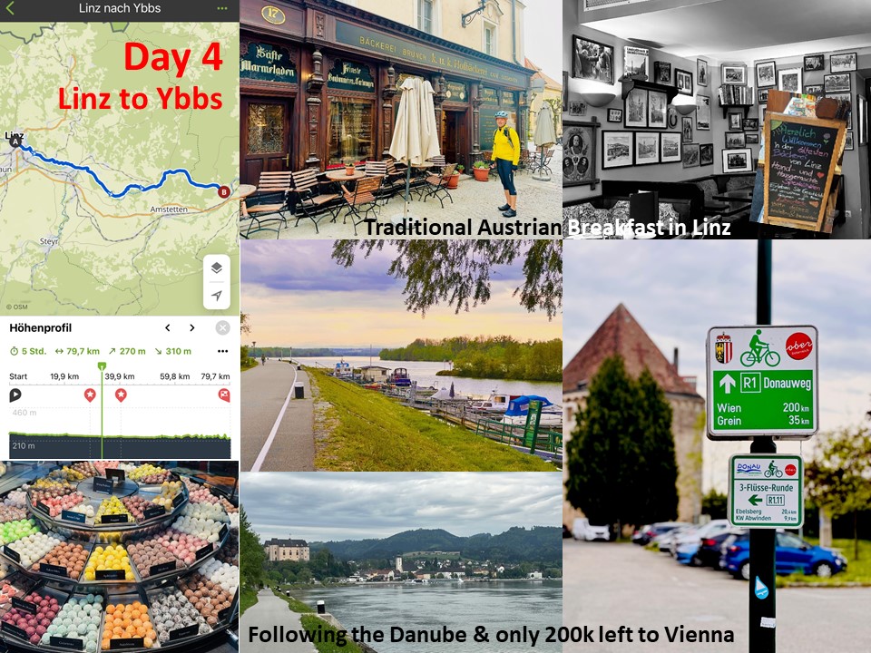
From Chondrometrics Headquarters in Freilassing to Messe Wien is a 350 km journey that typically takes around 3 ½ hours by car. However, by following the course of the 4 rivers Saalach, Salzach, Inn, and Donau (Danube), the distance extends to 450 km, requiring approximately 24 hours on a bicycle.
David Hunter from the University of Sydney and Felix Eckstein opted for the scenic route to the @OARSI Conference 2024, using the extended travel time to rejuvenate mind and body while enjoying informal discussions and camaraderie. Their expedition commenced from Freilassing to Burghausen on the first day, where the world's longest castle stands.

Day 1

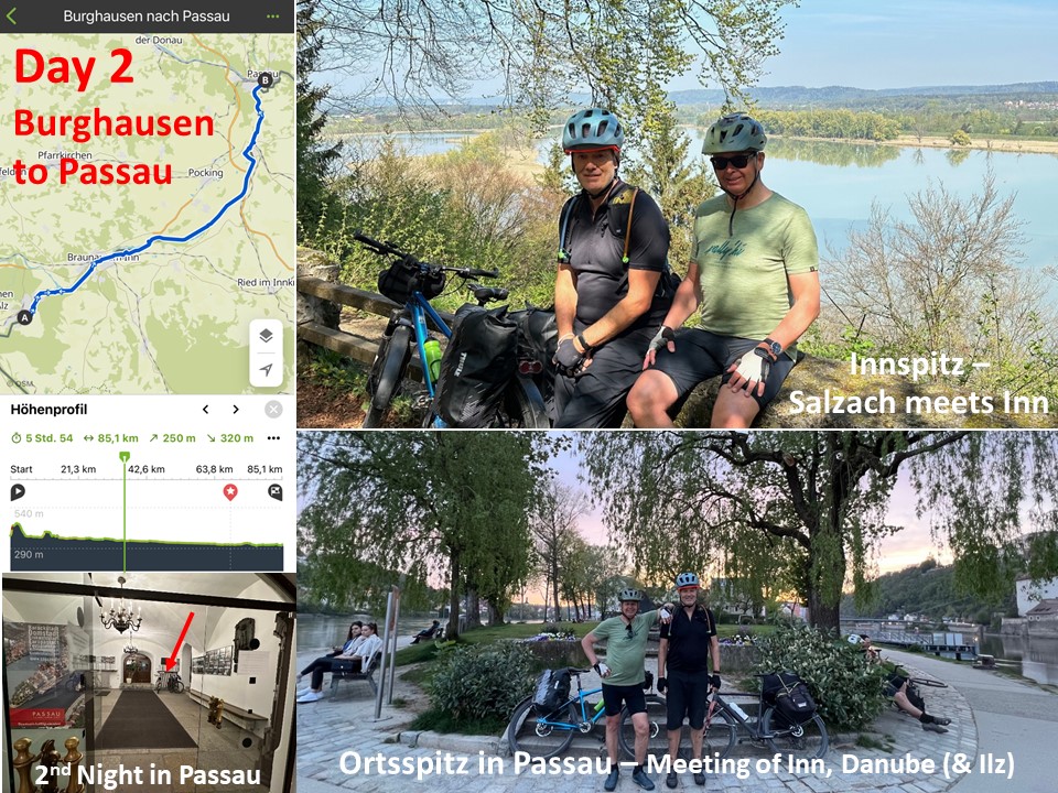
On the second day, they reached Passau, where the convergence of the Inn, Danube, and Ilz rivers often floods the historic town.  

Day 2

Continuing on the third day, they arrived in Linz, renowned for its blend of industry and artistry.

Day 3

The fourth day began with a hearty breakfast at the delightful Kaiser’s K&KBakery and concluded in the historic town of Ybbs after covering a modest 80 kilometers due to somewhat inclement weather.  

Day 4

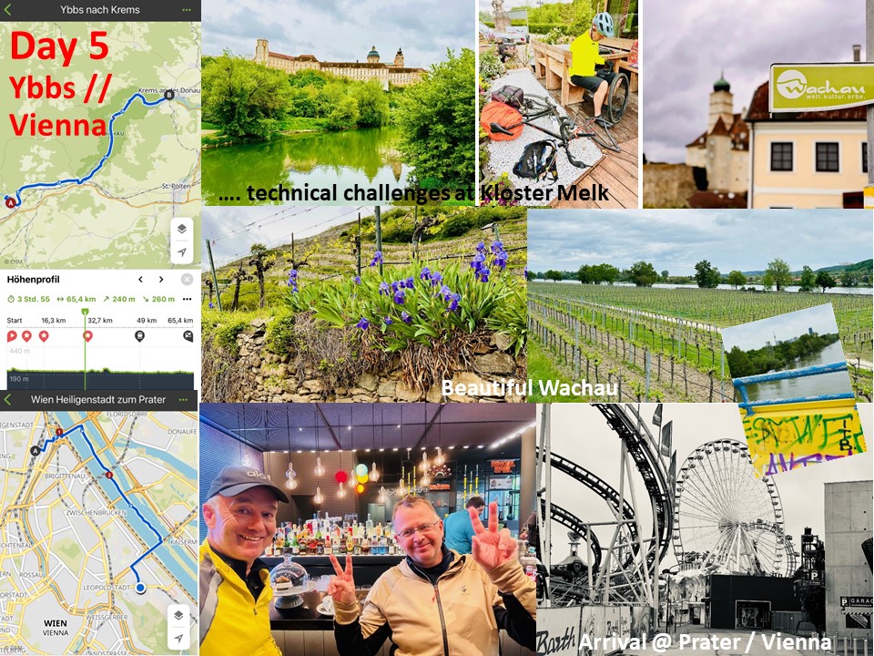
The following morning, both cyclists were greeted by the impressive Monastery of Melk, heralding their entry into the picturesque wine region of the Wachau. From the avant-garde town of Krems, they had to board a train to Wien-Heiligenstatt to ensure they stayed on schedule for their initial appointments in Vienna. Nonetheless, they still enjoyed a memorable "entrance" into Vienna by crossing the Danube_Island. Their legs and spirits alike rejoiced upon their arrival at Vienna / Prater, where an engaging OARSI conference awaited, promising to keep them occupied and inspired for the days ahead.

Day 5


EVENT
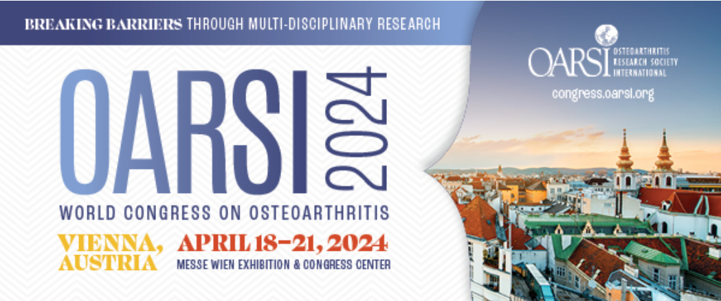
OARSI WORLD CONGRESS VIENNA, APRIL 18-21, 2024

Breaking Barriers Through Multi-Disciplinary Osteoarthritis Research - the OARSI World Congress in Vienna, April 18-21, 2024
Osteoarthritis and Cartilage

Scientists from Chondrometrics and PMU will be delivering an oral presentation and several posters on quantitative measurement methodology of articular tissue pathology, as featured in this post.

The OARSI World Congress is the largest global platform of those engaged in osteoarthritis (OA) research. It attracts professionals from diverse disciplines in academia & industry, basic, translational and clinical scientists, allied health professionals, and policy makers. This year, the congress is held at Messe Wien Congress Center, and anticipates attracting >1,000 attendees from >40 countries. OARSI, the Osteoarthritis Research Society International, is the premier society to advance the understanding and treatment of OA, a debilitating condition affecting >600 million people worldwide.

Felix Eckstein, Research Professor at Paracelsus Medical University (PMU, Salzburg, Austria), and CEO of Chondrometrics GmbH (Freilassing, Germany) has recently become the Austrian National Ambassador for OARSI. He enthusiastically anticipates the World Congress to be hosted in the country of his academic work, his research group being affiliated with the Ludwig Boltzmann Institute of Arthritis and Rehabilitation (LBIAR) under leadership of Tanja Stamm.

On Friday, April 19 (8:45-10:15, Abstract No. 020) he will give a talk supported by a team from Formation Bio, owner of a potentially disease-modifying OA drug: “Sprifermin”. The presentation will focus on whether analyzing magnetic resonance images (MRIs) without blinding to relative acquisition order affects observed effect sizes of drug efficacy on cartilage thickness in the FORWARD trial. He will also present a poster (No 90) on the clinical validation of fully automated measurement of cartilage composition using MRI (T2), employing deep learning (AI). This work involves researchers from Charité (Nicholas Brisson & Georg Duda) and 4P Pharma (Francis Berenbaum) and is part of both the OVERLOAD and OA-BIO Eurostars project.

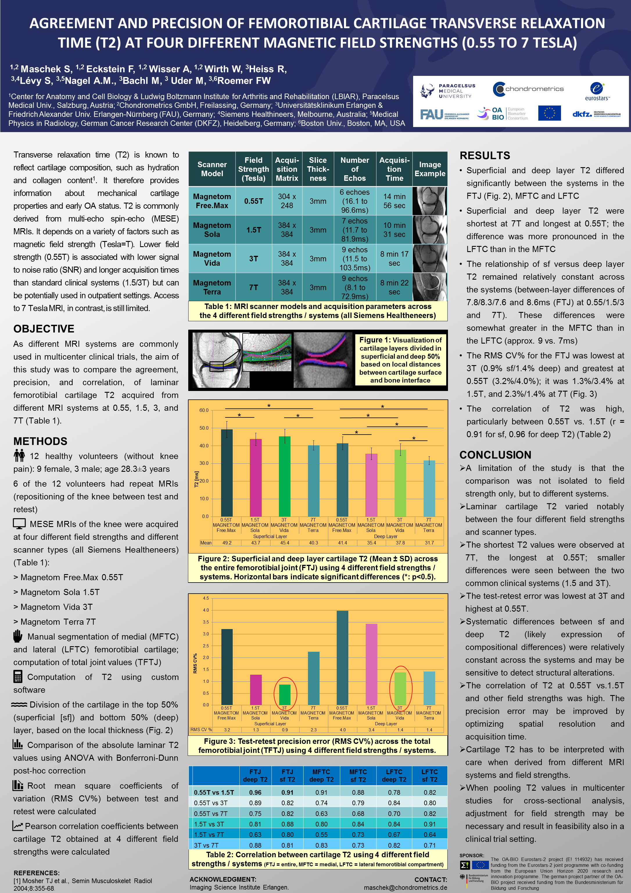
Susanne Maschek, Chief of Education and Quality Control at CHM, will present a poster (No 492) on the impact of MRI scanner field strength on cartilage T2. The study includes a low-field magnet (0.55 Tesla) suitable for outpatient settings, clinical ( 1.5 and 3.0T), and a high-field research scanner (7.0T). The work was performed in collaboration with Frank Roemer's group at the University of Erlangen and representatives of Siemens Healthcare.

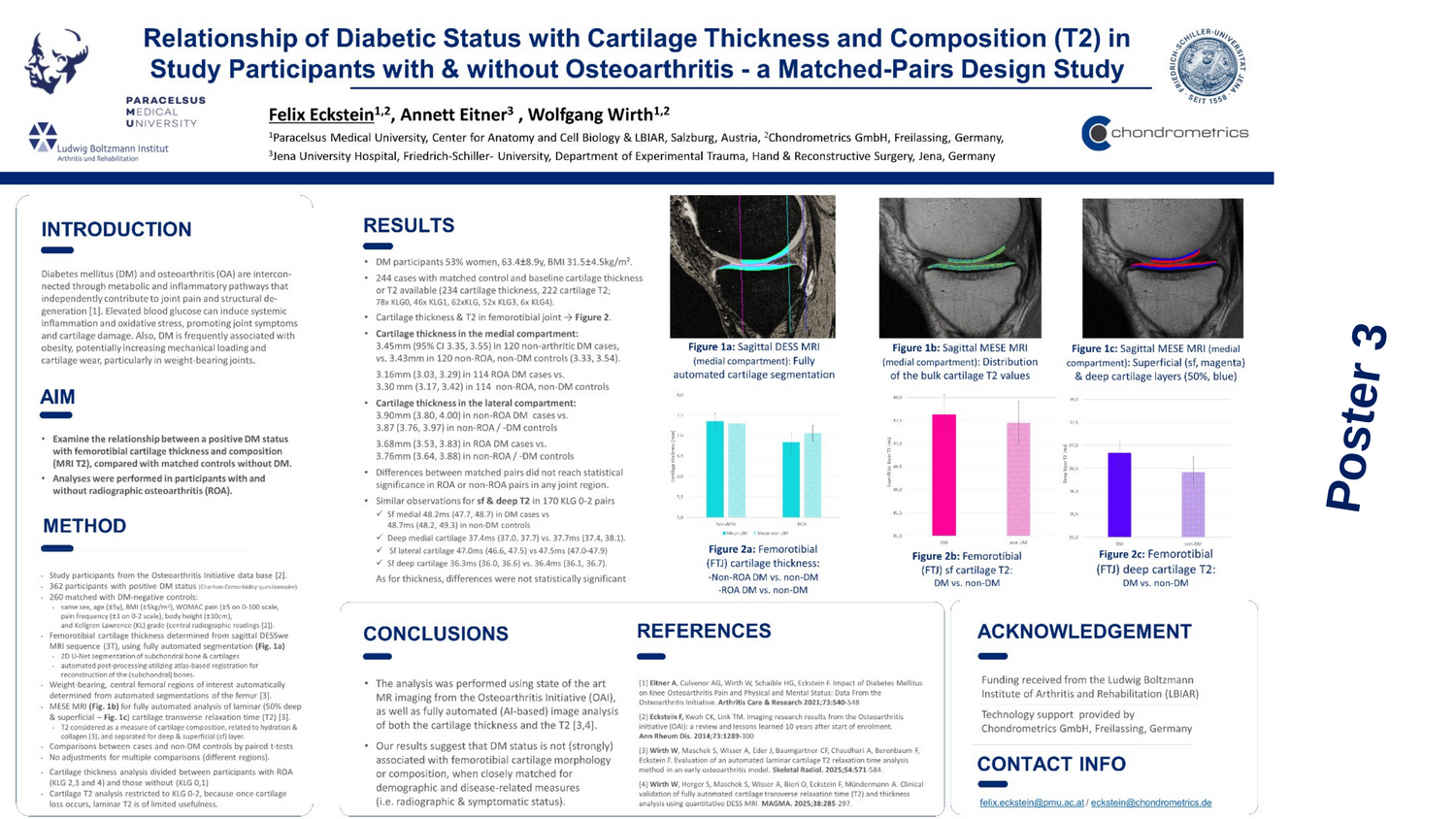
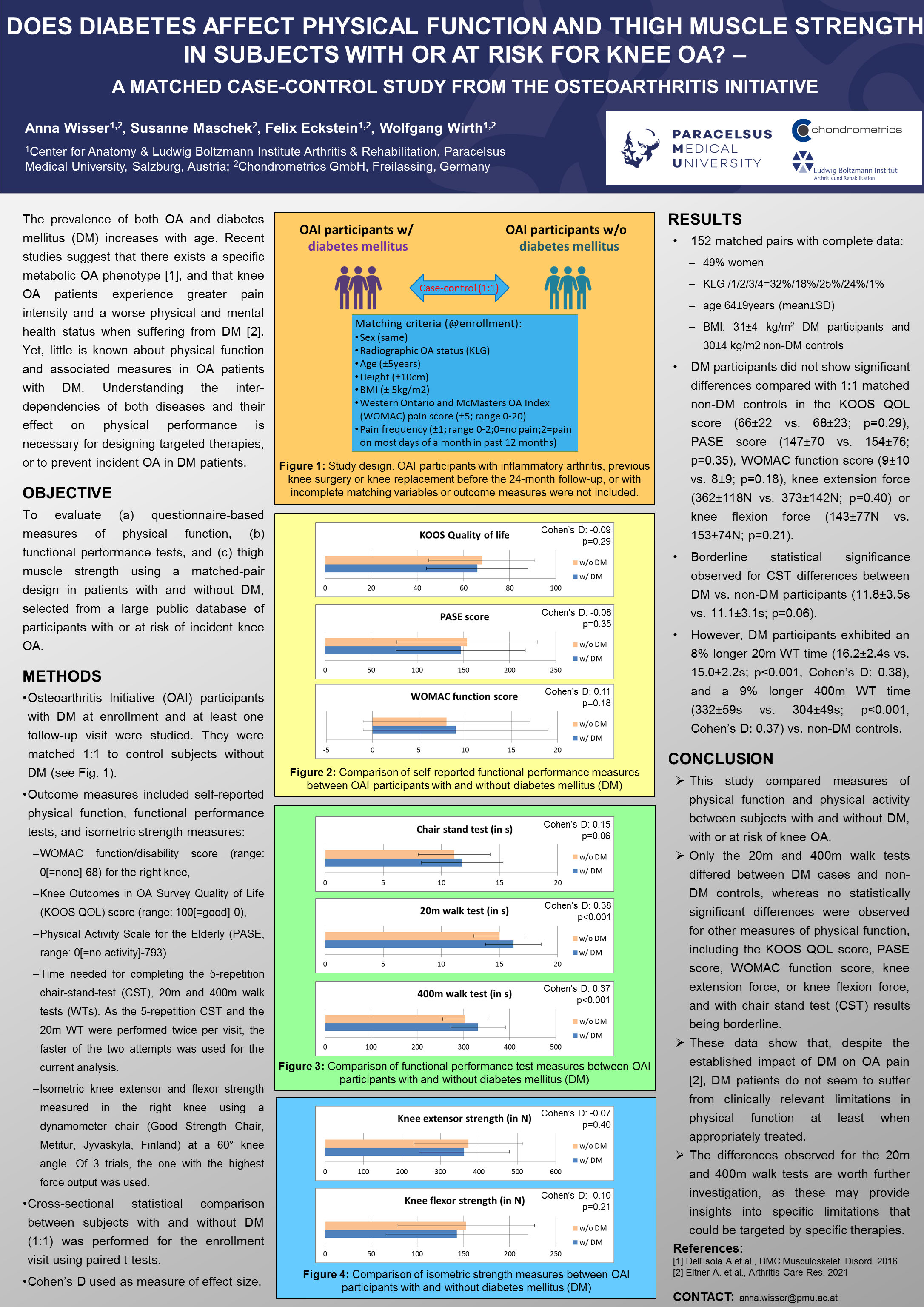
Anna Wisser, Project Manager at PMU and CHM, will present a poster (No 207) on whether diabetes mellitus affects physical function and thigh muscle strength in participants of the Osteoarthritis Initiative. The study is part of a broader project exploring the impact of blood surgar on tissue structure and pathology in OA within the LBIAR.

Clement Heistracher, PhD candidate of Jana Eder at PMU will contribute a poster (No 084) on fully unsupervised automated segmentation of muscle and adipose tissue of thigh MRIs. Automated segmentation is performed without labeled data, providing a substantial advance in automated image segmentation technology.

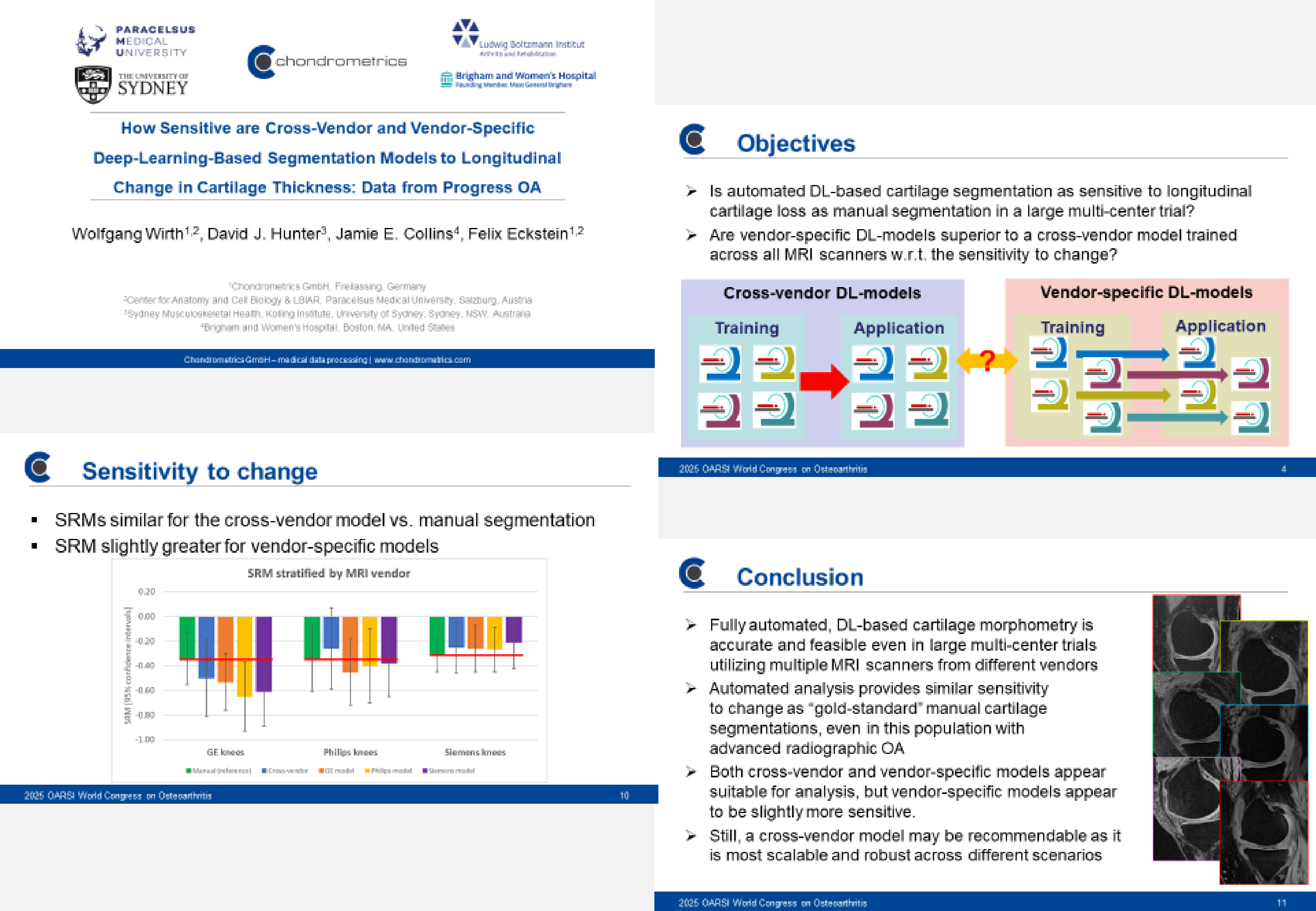
Wolfgang Wirth, Senior Postdoc at PMU and Chief Technology Officer at CHM, will present a poster (No 491) on fully automated segmentation, comparing the ability of different MRI sequences in detecting OA cartilage thickness change, with co-authors Akshay Chaudhari from Stanford University and David Hunter from the University of Sydney. A second poster (No 493) explores the magnitude of sex-differences in radiographic knee joint space width (JSW), a commonly used measure of OA status and progression. It further studies "genuine" sexual dimorphism in JSW and cartilage morphometric measuring, comparing female and male Osteoarthritis Initiative participants with the same body height. The poster is part of a broader initiative to characterize sex differences in (peri-) articular tissues as potential risk factors for knee OA, currently submitted to a special issue of the Journal of Osteoarthritis & Cartilage.


REVIEW ARTICLE
OSTEOARTHRITIS & CARTILAGE

30 Years of MRI-based cartilage & bone morphometry in knee osteoarthritis: From correlation to clinical trials
Osteoarthritis and Cartilage

It is with great pleasure and a bit of pride to see our review on 30 years of MRI-based cartilage and bone morphometry in knee osteoarthritis published in the special issue of Osteoarthritis and Cartilage Journal.

This exciting journey has taken us from initial “correlation”, i.e. method development with external reference standards for validation to exploring the physiology and pathophysiology of the disease, to qualification studies towards a surrogate endpoint, and finally to interventional “clinical trials”, exploring the structural efficacy of potentially disease modifying OA drugs (DMOADs) on articular tissues.

It has been a great fortune to be able to work on this review with our co-authors from Monash University in Melbourne (Australia): Anita Wluka and Flavia Cicuttini, and receive support of esteemed colleagues, namely Mylène Jansen (Universitair Medisch Centrum Utrecht), Alan Brett (Stryker - Imorphics) and Christoph Ladel (CHL4special consulting).

We are grateful for all the support we received and hope you enjoy reading the article

Felix Eckstein, Wolfgang Wirth
LinkedIn

EVENT
SCIENTIFIC ADVISORY BOARD MEETING
FREILASSING/SALZBURG, FEBRUARY 22-26, 2024

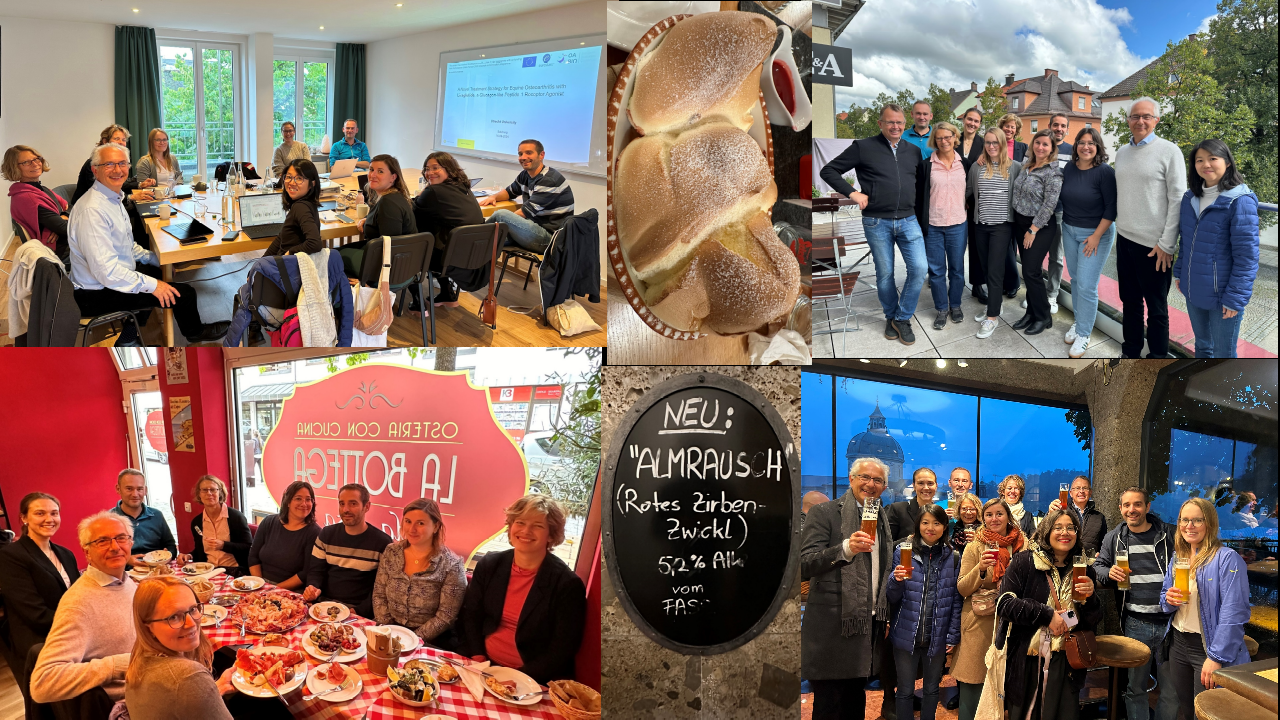
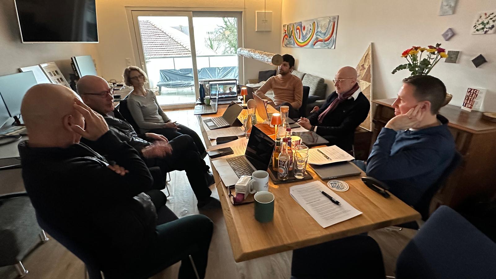
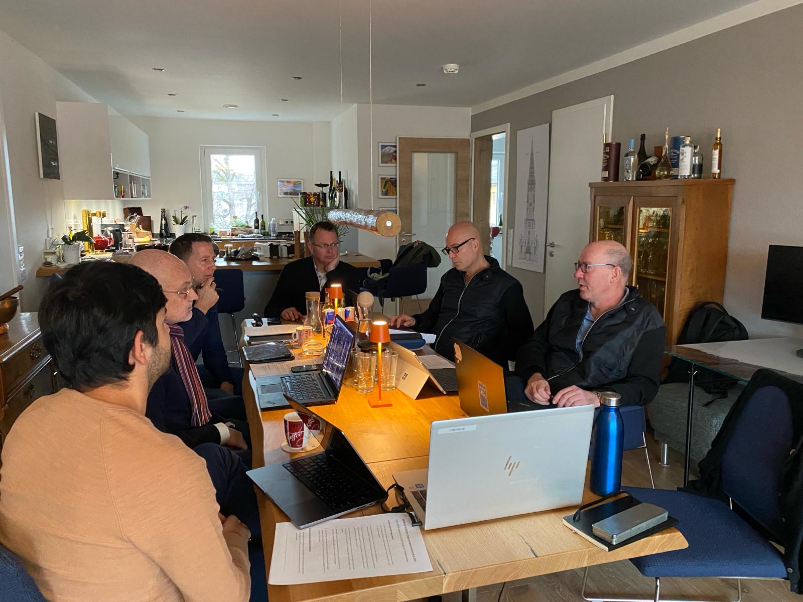
After initial meetings in 2019 & 2020, Chondrometrics organized the 3rd SABM in February 2024, now in the new office in Freilassing, and of course on the ski slopes of the Salzburger Land. 

Despite Lufthansa strikes causing turbulence with arrivals, everyone eventually reached Freilassing safely, albeit with some delays. Neal Bangerter (Imperial College), Mikael Boesen (University of Copenhagen), Akshay Chaudhari (Stanford University), Chris Ladel (4Consulting Darmstadt), and Ali Mobasheri (University of Oulu) turned out to be an incredibly productive and inspiring group. 

Advisory Board OfficeAdvisory Board Office 2

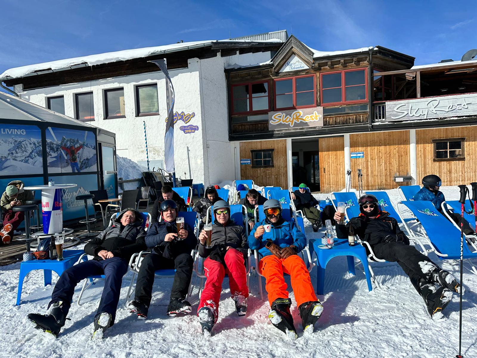
Topics for discussion included state-of-the-art and future MRI sequences, automated tissue segmentation using AI- and deep-learning based methods, quantitative methodology for analysis of synovitis, set-up of an “ideal” clinical trial for structure modification in osteoarthritis, and the acceptance of automated measurement methodology and imaging assessments as surrogate endpoints by regulatory agencies. Yet, as in former years, the group was also a lot of fun and enjoyed good company, friendship, restaurants, and great skiing at Leogang, Kitzbühl, and Fieberbrunn ski stations. Something to be certainly repeated.

Advisory Board RelaxingAdvisory Board Relaxing II

SUMMARY & PRESENTATIONS
INTERNATIONAL WORKSHOP OF OSTEOARTHRITIS IMAGING (IWOAI), LAUSANNE, JUNE 28-30, 2023

The team of Chondrometrics GmbH and the Research Program for Imaging at Paracelsus Medical University presenting at the Imaging Workshop in Lausanne

The team of Chondrometrics GmbH and the Research Program for Imaging at Paracelsus Medical University presenting at the Imaging Workshop in Lausanne included Susanne Maschek (Chief of Human Resources and Quality Control), Anna Wisser (Project Manager & PhD), Wolfgang Wirth (Chief Technical Officer and Chief of Quality Control), and Felix Eckstein (CEO & CMO).

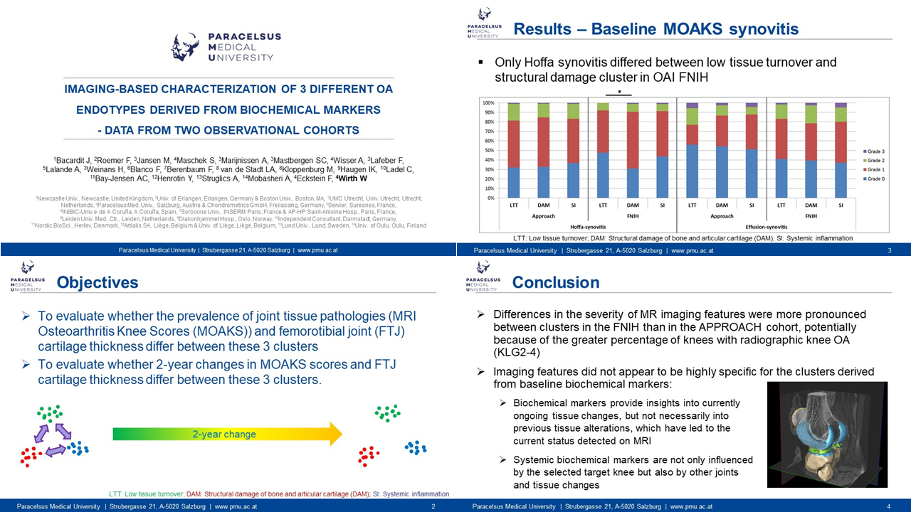
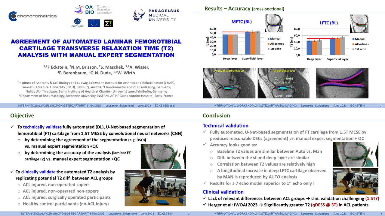
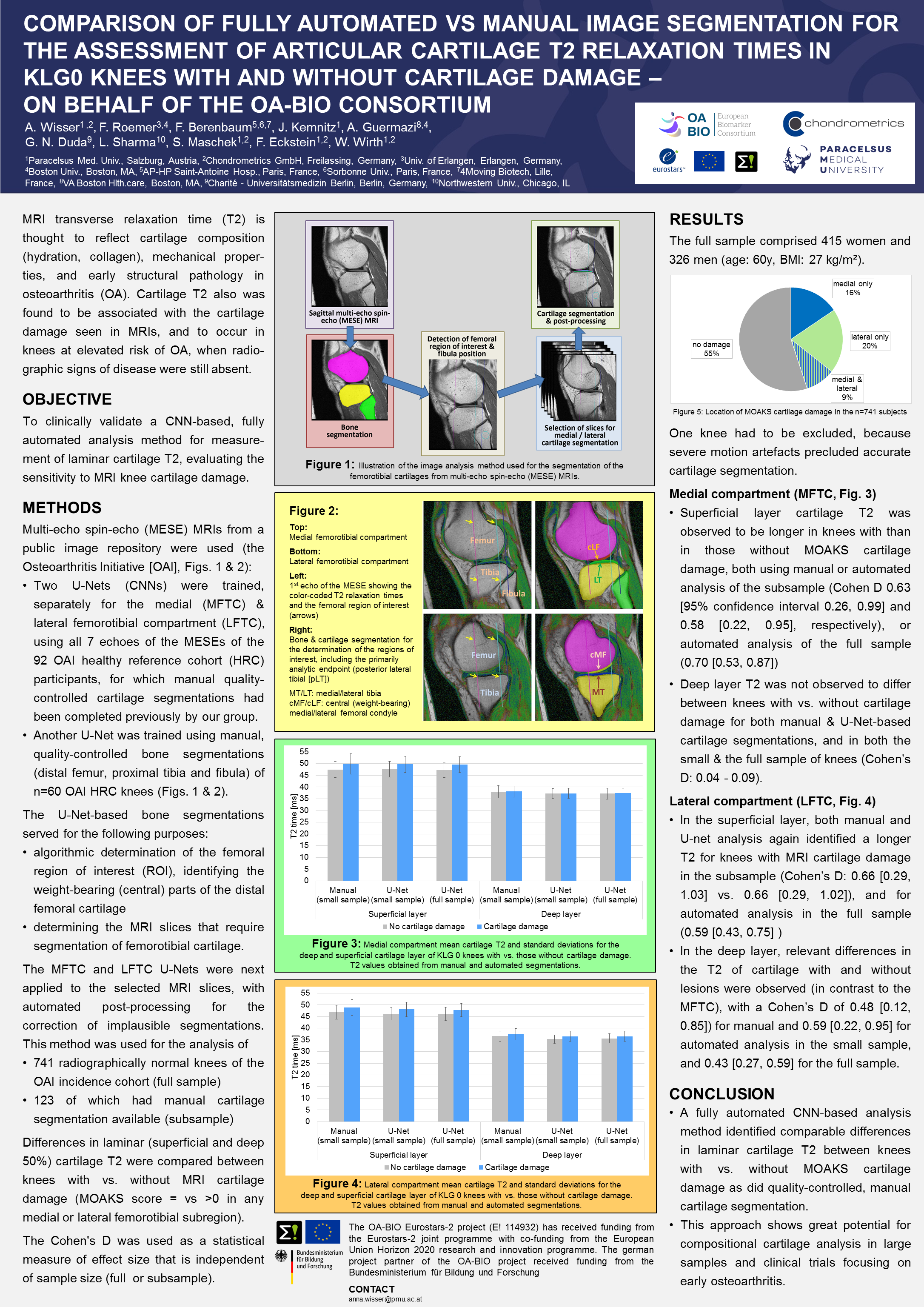
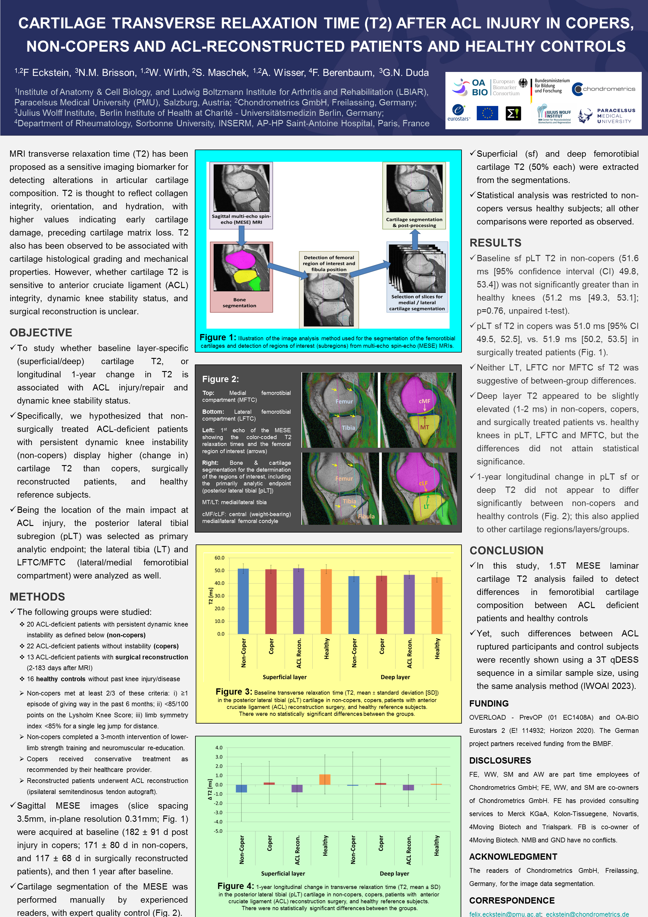
The group contributed 3 first author oral presentations and 2 posters displayed below. Susanne Maschek spoke on the variability of cartilage T2 measurements between MRI field strengths ranging from 0.55 to 7.0 Tesla, Wolfgang Wirth on the imaging characteristics of 3 different osteoarthritis endotypes derived from liquid (biochemical) markers, and Felix Eckstein on the agreement of laminar (deep and superficial) T2 between fully automated and expert manual analysis. Anna Wisser presented a poster on the agreement of automated vs. manual T2 analysis in radiographically normal knees with and without MRI cartilage lesions, and Felix Eckstein one on cartilage T2 in anterior cruciate ligament (ACL) injured patients (non-copers, copers, and operated) vs. healthy controls.

We thank our co-authors for the great collaboration on the above projects, and we look forward seeing you at the OARSI World Congress 2024 in April in Vienna as well as at the IWOAI 2024 in June in Marrakech.


First-author posters


EVENT
20thANNIVERSARY OF CHONDROMETRICS FREILASSING, APRIL, 2023

In April 2004, Chondrometrics was founded in Munich, Germany, as a spin-off of the Ludwig Maximilians Universität (LMU) München by Prof. Reinhard Putz and PD Felix Eckstein. At that time, no one would have thought that this was the beginning of a >20-year history. Reason to celebrate with the team and former team members
CHM 20th Anniversary Team 1

Chondrometrics was founded in Munich, Germany in April 2004, by Prof. Reinhard Putz and PD Felix Eckstein. What was initially thought to be an investment for a single project, turned out to be a >20 year success story that will hopefully continue for many more years. Thanks to wonderful colleagues and employees, the small University group, with initial experience in cartilage segmentation, emerged to a leading vendor of medial image analysis. Today, Chondrometrics participates in many important clinical trials of disease modifying osteoarthritis drugs (DMOADs), using its ISO 13485 approved quality management system and it’s under the current MDR “CE” approved software platform.

CHM 20th Anniversary Team 2

Big reason to celebrate! On April 22nd/23rd, two weeks after the actual date, the Chondrometrics team joined in Freilassing, the site of the new office. Although officially the location business since 2021, the 20th anniversary gathering also marked official inauguration of the premises. 

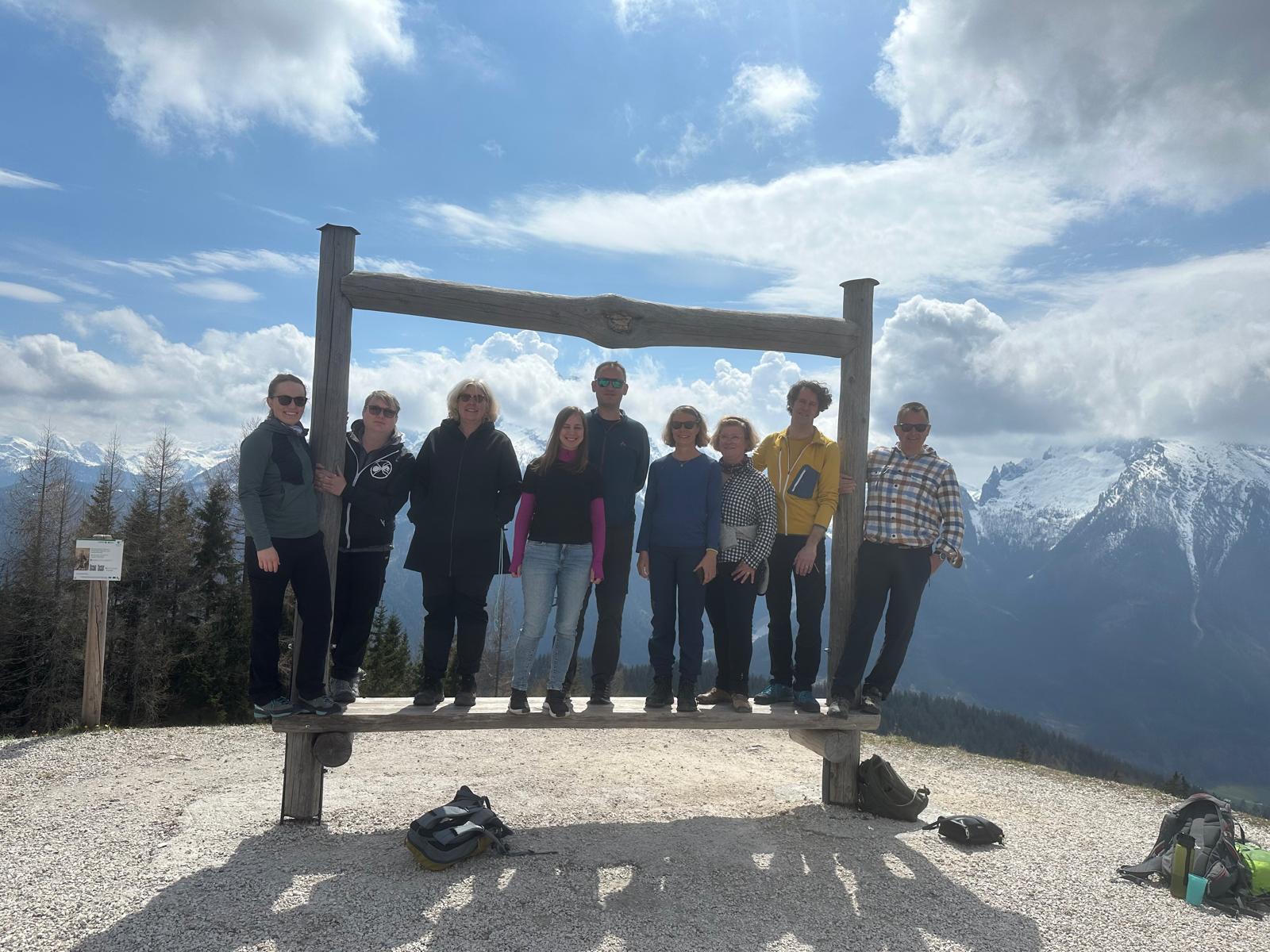
The team was joined by Prof. Reinhard Putz and former team members Jana Eder (geb. Kemnitz), Manuela Kunz, Jan Hohe, Tobias Stammberger, and David Fuerst. After “aperitivo” at the office, the party spent a nice Bavarian evening at the Zollhäusl, marking the close border to Austria, and then went hiking on the next day to the Hochschwarzeck, where lunch was taken in the sunshine with a splendid view onto the mountain peaks of the Chiemgau.

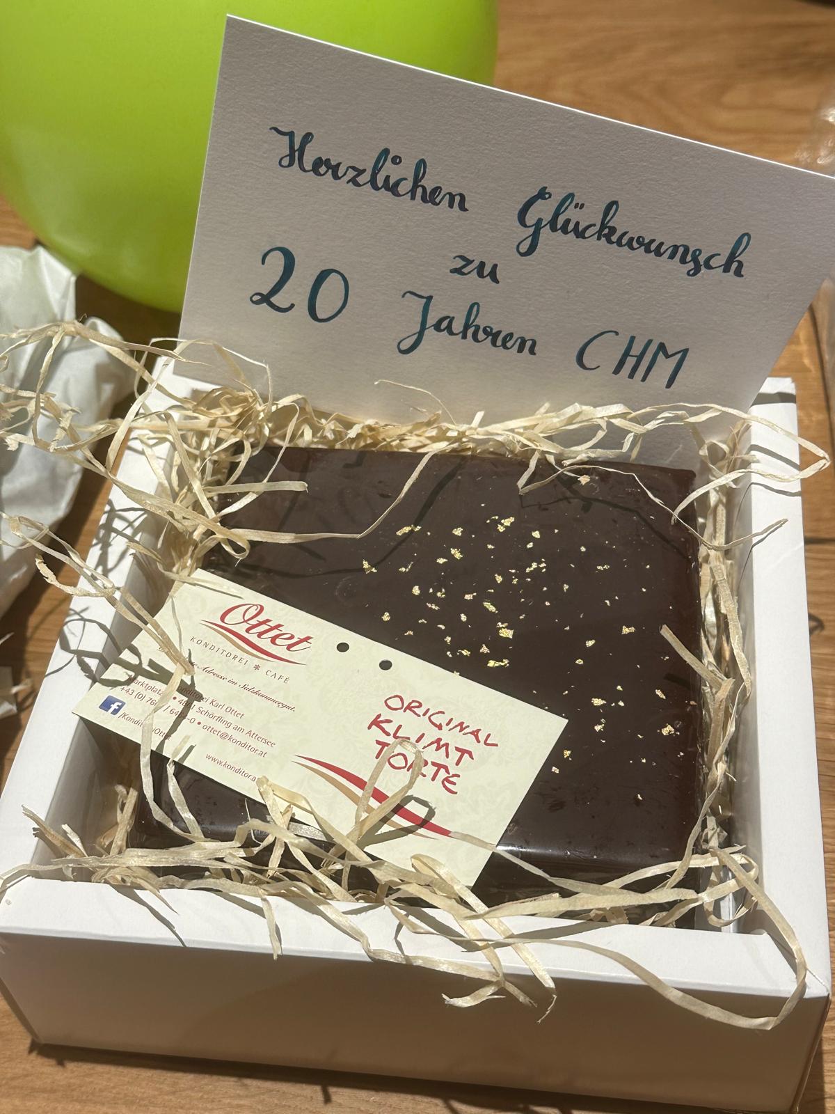
CHM 20th Anniversary Cake

Plans are already being made for the 25th anniversary, in April 2028


SUMMARY & POSTERS
OARSI WORLD CONGRESS, DENVER, MARCH 15-19, 2023

The team of Chondrometrics GmbH and the Research Program for Imaging at Paracelsus Medizinische Privatuniversität presenting at the World Congress in DENVER

The team of Chondrometrics GmbH and the Research Program for Imaging at Paracelsus Medizinische Privatuniversität presenting at the World Congress in Denver included  Wolfgang Wirth (Chief Technical Officer and Chief of Quality Control), Anna Wisser (Project Manager), Kalpana Sharma (PhD student), and Felix Eckstein (CEO & CMO).
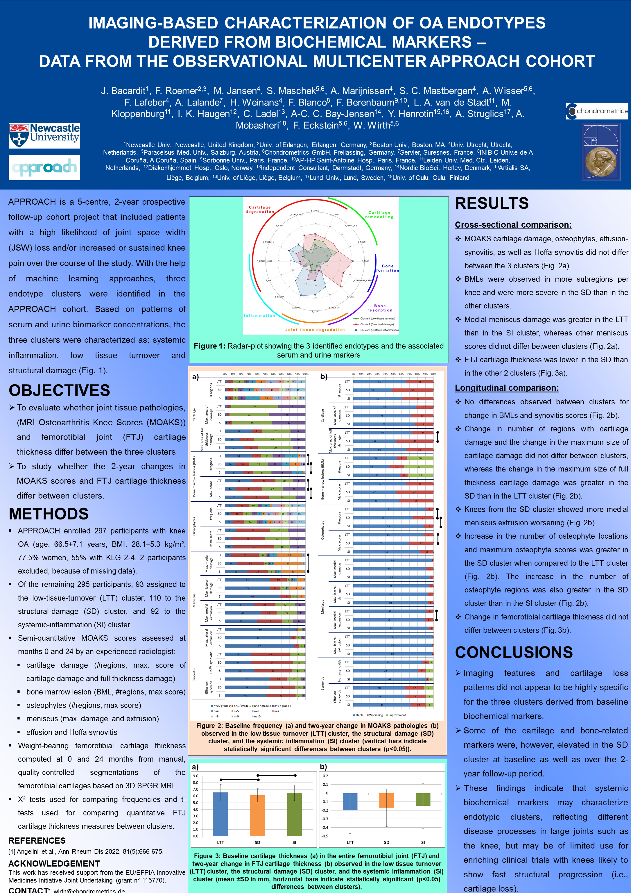
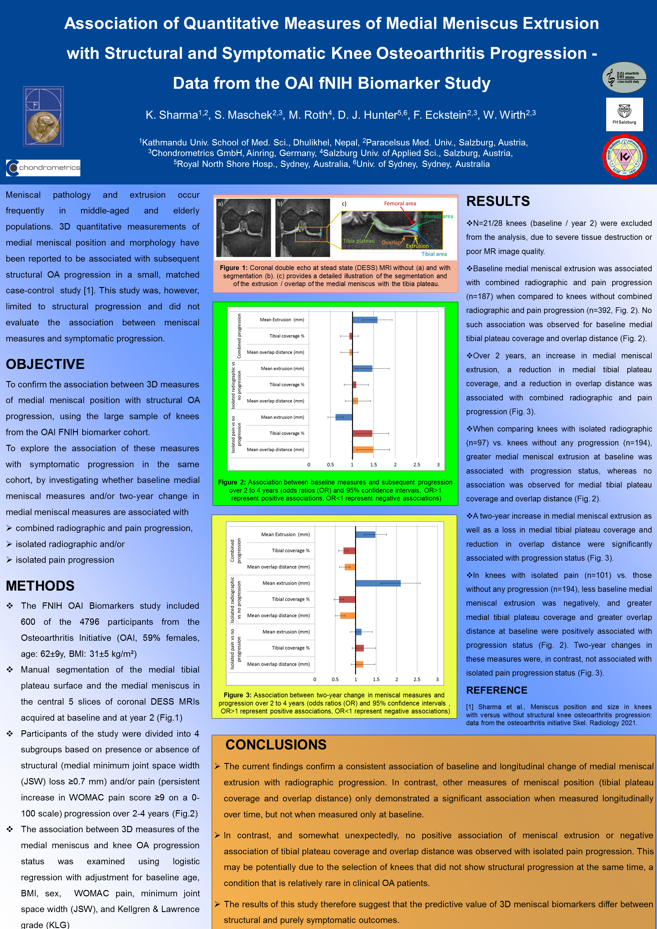
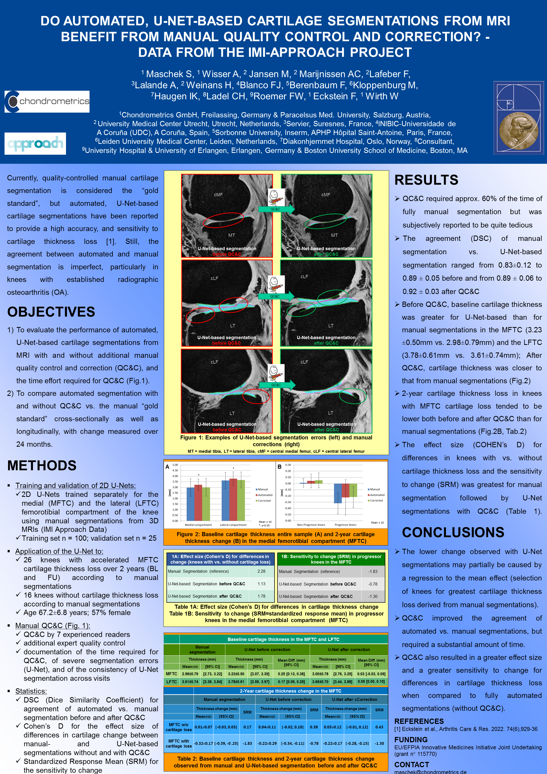
The group presented the 5 first-author posters displayed below. These reflect the current project status at Chondrometrics and at PMU. They cover topics ranging from Imaging-based characterization of osteoarthritis endotypes (APPROACH project), the relationship of meniscus extrusion with structural and symptomatic osteoarthritis progression (OAI FNIH 1 Biomarker Consortium project), the clinical validation of automated (U-net-based) cartilage T2 analysis using two different clinical models (Eurostars OA BIO project), and finally a project studying whether automated cartilage thickness analysis profits from quality control and correction compared with manual gold standard segmentations.

We would like to thank our many collaborators for their support and contribution to the above projects, and it will be a pleasure seeing you all at the 2023 Imaging Workshop in Lausanne


First-author posters


TEAM & SERVICES

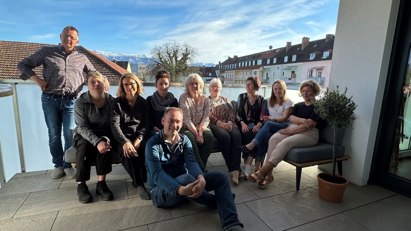
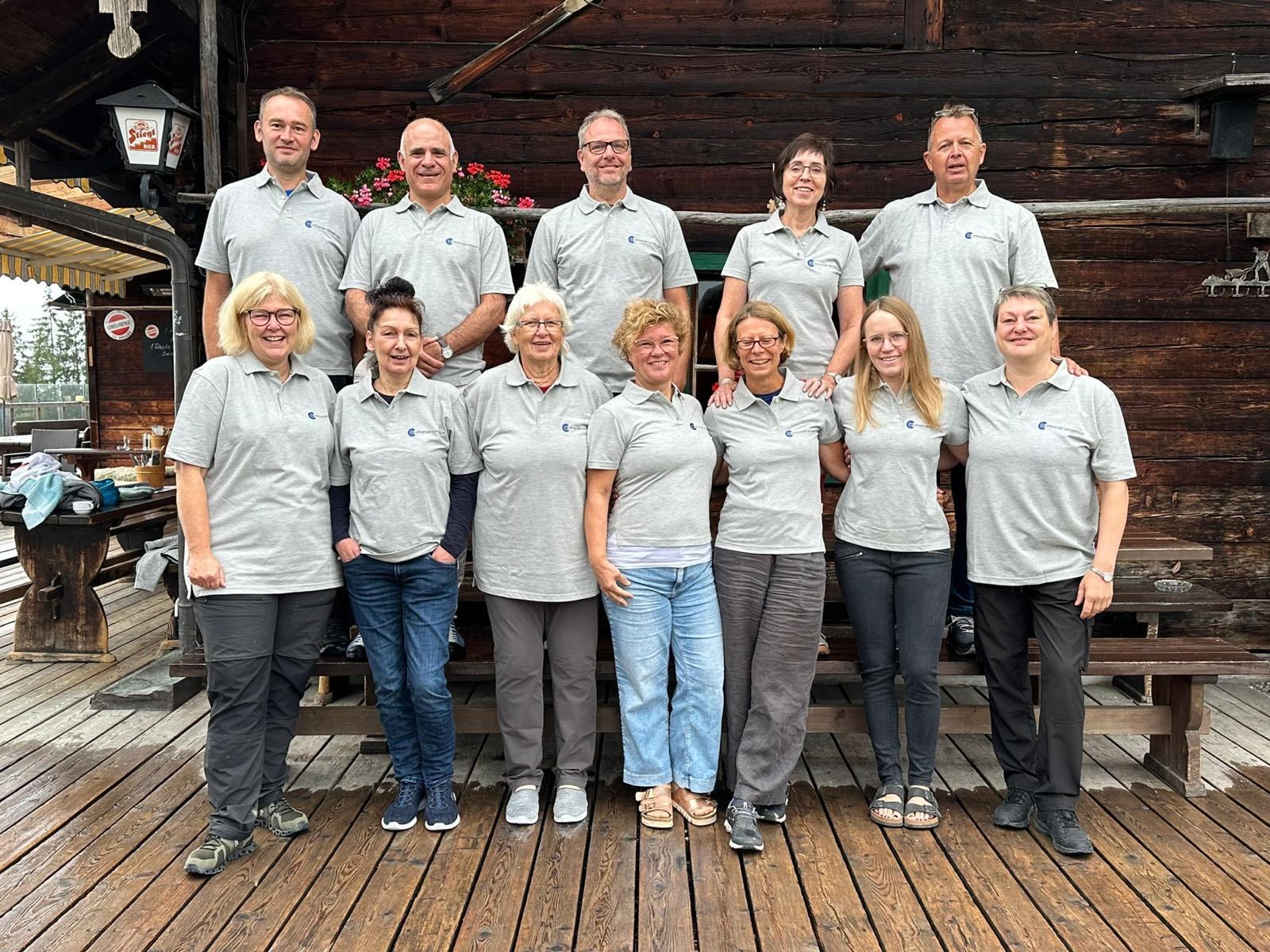
Chondrometrics TeamChondrometrics GmbH provides expert consultation and image analysis service to academic researchers and the pharmaceutical industry. The strength of Chondrometrics, we believe, lies in the dedication and experience of its team of specialized researchers and readers, the profound integration with the scientific community, the rigorous scientific validation of its methodologies, and the transparency of its technology by publication in leading scientific journals. At Chondrometrics, we value our clients as collaborators, react immediately to their requests and specific needs, provide validated scientific information, communicate promptly, and act with anticipation.

The service of Chondrometrics supports:

  • Clinical studies, investigating the efficacy of disease modifying osteoarthritis drugs (DMOADs) of other therapeutic approaches in osteoarthritis
  • Animal studies, investigating the efficacy of DMOADs and other therapeutic approaches in OA
  • Epidemiological studies, investigating the risk factors of OA incidence and progression
  • Basic research studies, investigating functional adaptation and the biomechanics or articular tissues
  • Technical development of image analysis software solutions for quantitative measurement of anatomical structures, including strategies for validating these measurements

The service of Chondrometrics involves:

  • Consultation on study designs and MR image acquisition protocols
  • Quality checks or continuous monitoring of image acquisitions in multicenter studies
  • Segmentation of relevant anatomical structures in medical image data by its team of well-trained and highly experienced readers
  • Quality controls of all segmented images by a team of experts
  • Computation of 3D quantitative endpoints, including total joint structures and anatomically defined subregions
  • Delivery of quantitative endpoints to customer database in requested format
  • Consultation on interpretations of study results

The team of Chondrometrics includes:

Operational Management
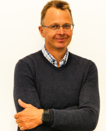
  • Prof. Dr. Felix Eckstein, MD, Chief Executive Officer & Chief Medical Officer,
    Felix Eckstein

    Felix Eckstein

    Current Position: CEO and CMO at Chondrometrics GmbH
    Mail to: eckstein@condrometrics@de
    Mobile: +49 162 7316401
    Degree: Univ. Professor, Dr. med. (MD)

    Felix Eckstein was born in Freiburg, Germany. He studied Medicine in Freiburg and Heidelberg to graduate in 1991. He received a scholarship from the German Academic Exchange Service (DAAD) to study Medicine at Bristol University (U.K.) in 1987/1988 and a scholarship from the Dr. Carl Duisberg Stiftung to complete a doctoral thesis at the University of Innsbruck (Austria) in 1988/1989.

    In 1991, Felix Eckstein joined the research group of Prof. Reinhard Putz at the Institute of Anatomy at the Ludwig-Maximilians-University (LMU), Munich. In 1993, he joined the Institute of Diagnostic Imaging at LMU and started working on the quantitative analysis of articular cartilage from MRI. Back to Anatomy in 1995, he completed his „Habilitation“ at the Institute of Anatomy in 1997. 

    In 2003, he founded Chondrometrics GmbH, to become a leading provider of medial image analysis services to researchers in academia and the pharmaceutical industry. 

    In 2004, Felix Eckstein became Professor of Anatomy and Director of the Institute of Anatomy at Paracelsus Medical University (PMU) in Salzburg. His research focuses on the integration of imaging methods for understanding the morphology, function and pathophysiology of musculoskeletal tissues, in particular osteoarthritis and osteoporosis.

    In 2019 he assumed a research professorship to became Head of the “Research Program for Musculoskeletal Imaging “at the Center of Anatomy & Cell Biology at PMU. He continues working on imaging biomarker qualification for the use as surrogate endpoints in clinical trials on disease modifying therapies He also focusses on the clinical validation of automated measurement technology, based on AI- and deep learning (DL) approaches.

    He organized the 1st Int. Workshop on Osteoarthritis Imaging (IWOAI) in Ainring in 2007, the 5th/14th IWOAI in Salzburg in 2011/2020, and the Annual Meeting of the Anatomical Society in Salzburg in 2014.  

    Felix Eckstein was president of the German Society of Biomechanics (DGfB) in 2002/3, Secretary General of the Osteoarthritis Research Society International (OARSI) in 2005/6, board member of OARSI in 2005-2009, and was invited to be a Member of the German National Academy of Science (Leopoldina) in 2013.

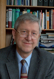
  • Prof. Dr. Reinhard Putz, MD, Vice President,
    Reinhard Putz

    Reinhard Putz

    Current Position: Vice president
    Email: reinhard.putz@med.uni-muenchen.de
    Degree: Univ.-Prof., Dr. univ. med. (MD)

    Reinhard Putz was born in Innsbruck/Austria. He studyied medicine and graduated in 1968. He became Assistant Professor at the University of Innsbruck Institute of Anatomy, where he completed his postdoctoral thesis (Habilitation) in 1979. After research stays in Munich and Freiburg/Germany, Professor Putz became Director of the Institute of Anatomy at Albert-Ludwigs-Universität Freiburg. In 1989, Professor Putz was appointed Director of the Institute of Anatomy at Ludwig-Maximilians-Universität (LMU) München. Between 2003 and 2010 he served Vice- President of LMU. Professor Putz retired from his academic positions in September 2010. In 2003, Professor Putz was a cofounder of Chondrometrics GmbH and since serves as Vice President of the company.

    Professor Putz’s research focuses on the biomechanics of the skeletal system, especially the formfunction relationship of the joints and the spine. He has also addressed various topics concerning clinical anatomy and general aspects of medical education, leading to approximately 250 publications and more than oral 450 presentations. Professor Putz was instrumental in producing a series of anatomy textbooks with international circulation (e.g. Sobotta, Atlas of Anatomy). Reinhard Putz was engaged in a series of scientific journals and serves as Associate Editor of Advances in Anatomy, Histology and Embryology.

    Reinhard Putz was president of the European Association of Clinical Anatomy (EACA) form 1991 to 1993, Associate Secretary General of EACA from 1993 to 2003, Vice-President of the German Society of Biomechanics (DGfB) 1997-1999, member of the board of the “Anatomische Gesellschaft” from 1998 to 2002, member of the board of the German Reunion of Medical Schools from 2003 to 2006. He was a reviewer of the German Research Foundation from 1996 to 2004 and member of the Board of National Medical Examination (part 1) from 1993 to 2003. Since 2009 Reinhard Putz is member of the board of governors of Medical University of Innsbruck/Austria.

    The academic work of Professor Reinhard Putz has been recognized in the form of numerous national and international awards and prizes. These include an honorary degree from the University of Constanta (Romania) and membership in the German National Academy of Sciences Leopoldina and the European Academy of Sciences and Arts. He also received the Friedrich-Pauwels Medal of the German Society of Orthopedics, and the Ars Legendi award by the German Rectors’ Conference and Donor’s Association for German Science for “Excellence in teaching“ and for organising a new master program (“Master of Medical Education”) together with the University of Heidelberg.

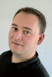
  • Dr. Wolfgang Wirth, PhD, Chief Technology Officer and Quality Manager,
    Wolfgang Wirth

    Wolfgang Wirth

    Current Position: CTO and Quality Manager
    Email: wirth@chondrometrics.de
    Mobile: +49 175 16 16 561
    Degree: Dr. rer. biol. hum Dipl.-Inf.

    Wolfgang Wirth was born in Fürstenfeldbruck, German. He studied Computer Science at the Technical University of Munich (TUM) and graduated in 2006. While studying at the TUM, Wolfgang Wirth was a co-founder of BitoS GmbH in 2004, a company focusing on mobile applications. In 2003, Wolfgang Wirth became a free-lancer at Chondrometrics GmbH, for which he develops software applications for quality control, segmentation and quantitative analysis of anatomical structures, including cartilage, meniscus and muscle.

    He completed his master thesis titled “Automatic detection of subregions in cartilage plates for the quantitative analysis in osteoarthritis patients” and graduated with a “Master of Science” degree in “Computer Science” from TUM in 2006.

    From 2007, Wolfgang Wirth worked at the Institute of Anatomy of the Ludwig-Maximilians-University (LMU) in Munich, Germany in the research group led by Prof. Reinhard Putz. Professor Putz also was supervisor for his PhD doctoral thesis titled “Longitudinal Analysis of Cartilage Morphology in Subregions of Knee Osteoarthritis Patients”.

    Since 2009, Wolfgang Wirth works at the “Institute of Anatomy and Musculoskeletal Research” at Paracelsus Medical University (PMU) in Salzburg, Austria, led by Prof. Felix Eckstein. In 2010, Wolfgang Wirth became co-owner of Chondrometrics GmbH and is responsible for the IT infrastructure, software development, and quality management in the company. Wolfgang Wirth has authored and co-authored more than 100 original scientific papers in leading scientific journals.

  • Dr. Susanne Maschek, PhD, Chief of Human Resources, Education, and Quality Control,
    Susanne Maschek

    Susanne Maschek

    Current Position: Chief of Human Resources, Education, and Quality Control
    Email:maschek@chondrometrics.de
    Mobile: +49 177 4458099
    Degree: Dr. med. vet. (PhD)

    Susanne Maschek was born in Munich, Germany. She studied Veterinary Medicine at the LMU Munich, Germany and graduated in 1995.

    From 1995 to 1997 Susanne Maschek worked with the group of Prof. Fritz Grimm at the Institute of Avian Medicine, LMU, Oberschleißheim, Germany for her PhD doktoral thesis titled "Studies of Heavy Metal Toxification in Predatory Birds, and Multi-Element Analyses Using Neutron Activation Analysis in Feathers". She performed the neutron activation analysis at the Research Reactor Center of the Department of Physics, TUM, Garching under the guidance of Prof. Schreckenbach, where she had a part time employment.Susanne Maschek worked also as substitute in several veterinary surgeries during this period.

    From 1999 on she was employed in a veterinary surgery, Oberaichbach, Germany, with focus on tall animals (cattle, horses) and birds until her child-raising leave (2001 to 2003).

    From 2004 to 2014 Susanne Maschek worked as freelancer at Chondrometrics GmbH. In 2015 she became Chief of Human Resources, Education, and Quality Control. She performs segmentations and is responsible for supervision and quality control of quantitative readings, as well as for the training and education of technicians. Since 2010 she is co-owner of Chondrometrics GmbH.

    Susanne Maschek also performs segmentation and quality control readings freelance-based for the Institute of Anatomy and Cell Biology of the Paracelsus Medical University (PMU) in Salzburg, Austria.

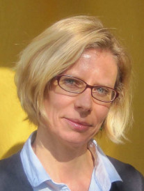
  • Anna Wisser, M.Sc., Project Manager
    Anna Wisser

    Anna Wisser

    Current Position: Project Manager
    Email: wisser@chondrometrics.de
    Mobile: +49 178 2056552
    Degree: M.Sc. Human Technology in Sports and Medicine

    Anna Wisser was born in Böblingen, Germany. She studied "Sport Science" and "Sport Therapy" at the University of Freiburg and graduated in 2014. Following her Bachelors degree, she continued her education in the master program "Human Technology in Sports and Medicine" at the German Sport University Cologne. She completed the degree with her thesis on the "Validation of an IMU-based Measurement System in Preparation for Clinical Gait Analysis" in 2017.

    During her studies she gained in-depth knowledge in the core areas of sport science, as physiology and biomechanics, as well as in fields like data management and analysis. Outside of her education in the field of sport science she was educated in business administration, business strategy and project management.
    From September 2018 on Anna Wisser has been supporting the Chondrometrics team in the areas of project management and quality control. She is also enrolled in the Medical Science Doctorate Study Program (PhD) at the Paracelsus Medical University (PMU) in Salzburg. Her research project deals with the "Dependence of functional performance measures (chair stand and walk tests) on lower limb pain, radiographic osteoarthritis status, and thigh bone / muscle / adipose phenotypes".

    Software Development, Quality Management, Data Base Management
    • Dr. Wolfgang Wirth, PhD
    • Dr. Jan Hohe, PhD
        Project Management and Quality Control
        • Dr. Susanne Maschek, PhD
        • Anna Wisser, M.Sc.
        Training, Supervision, and Quality Control of Quantitative Readings
        • Dr. Susanne Maschek, PhD
        • Prof. Dr. Felix Eckstein, MD
        Readers (Segmentation)
        • Gudrun Goldmann
        • Susanne Maschek, PhD
        • Jana Daimer
        • Sabine Mühlsimer
        • Annette Thebis
        • Barbara Wehr, PhD

        RESEARCH

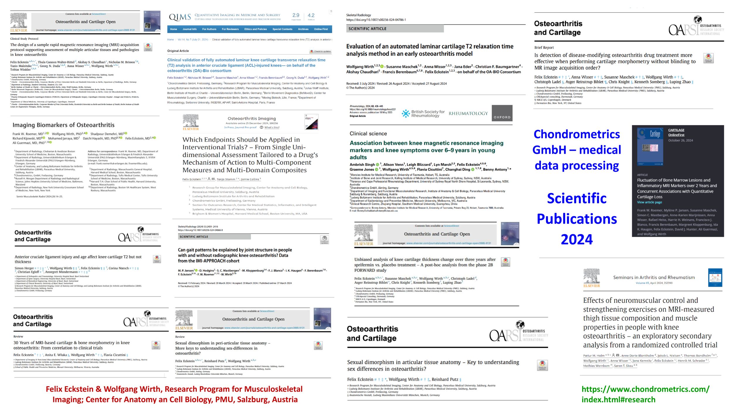
        Representatives of Chondrometrics are actively involved in academic research and publish continuously and extensively in the biomedical literature. The current section provides snapshots on these activities, presenting:

        1. Publication Indices for lead authors of Chondrometrics
        2. Exclusive Executive Summaries of published papers on key topics, including Pdfs
        3. Published articles by company authors listed by most cited or most current
        4. Grants, Subcontracts, and Service orders from academic sponsors
        5. A preset tool for searching Chondrometrics publications

        The Google Scholar publication metrics of Chondrometrics authors (Date: July 31st, 2024)

        Felix Eckstein All Since 2019
        Total Citations 37365 11715
        H-Index * 106 50
        i10-index ** 431 297
        Wolfgang Wirth All Since 2019
        Total Citations 7983 4045
        H-Index * 52 35
        i10-index ** 146 129

        * for explanation of this widely accepted metric of scientific impact, please check: https://de.wikipedia.org/wiki/H-Indexhttps://en.wikipedia.org/wiki/H-Index
        ** The i10 index is a specific metric used primarily within Google Scholar, to evaluate the “breadth” of scientific impact of a specific researcher. The index counts all authored or co-authored publications that are cited >10x since publication 

        In our Exclusive Executive Summaries section, we are synthesizing series of current publications around focus topics. These “mini-reviews” provide short overviews over key areas of interest, as well as archives for downloading PDFs of the relevant articles (click the reference# to download the pdf [when available open access]).
        Enjoy reading and do not hesitate to contact us for questions or further discussion.

        The Sprifermin Program

        Several Original Articles published 2013-2021 provide exciting insights into the structural benefits observed with Sprifermin in the Phase I, IIa, and IIb study.
        Spriferim Difference From Placebo

        The Sprifermin program is unique amongst potential disease modifying osteoarthritis drug (DMOAD) trials, as it encompasses several large multicenter randomized control trials, providing a tremendously rich resource for designing future DMOAD trials. With 5-year follow-up, FORWARD (Phase IIb) was the longest OA trial, using MR imaging, to date.
        The phase I “First in Man” (FIH) randomized control trial (RCT) studied the safety and potential efficacy of intra-articular sprifermin (rhFGF18) in 73 knee osteoarthritis (KOA) patients, scheduled for knee replacement. The study was suggestive of an anabolic effect on medial femorotibial cartilage, and of a reduction of knee replacement rates, without these observations reaching statistical significance (1).  

        In the following phase IIa “Proof of Concept” POC study, 192 KOA patients were randomized to placebo or 1 of 3 sprifermin doses, injected 3 times over 3 weeks, and the same set of injects again 3 months later (2). In this study, the dose-response in the primary structural endpoint (central medial femorotibial cartilage thickness) was distinguishable, but did not reach statistical significance, at months 3, 6 or 12 follow-up. However, rhFGF18 significantly increase cartilage thickness in the lateral and total (medial and lateral) femorotibial compartment relative to placebo-injected knees (2). There was a substantial reduction in knee pain in all study participants over 12 months, but no statistically significant effect of rhFGF18 in this study relative to placebo. Location-independent analysis of cartilage thickness change (see another Executive Summary on this Page) revealed that rhFGF18 not only increased cartilage thickness (at potentially non-useful locations in the joint), but effectively reduced cartilage loss (3).

        In the phase IIb study (FGF-18 Osteoarthritis Randomized Trial with Administration of Repeated Doses / FORWARD), 549 participants were randomized to 4 dose and 1 placebo group, with injection cycles at screening, 6,12, and 18 months (4). The patients included displayed Kellgren Lawrence Grade (KLG) 2-3 (medial or lateral disease), with a medial radiographic joint space width (JSW) of >2.5mm. This study found a statistically significant dose-dependent effect on the primary endpoint (total femorotibial cartilage thickness) as well as the central medial femorotibial subregion at year 2 and 3 (4), but no effect on medial radiographic JSW. Reduction in WOMAC pain (approximately 50%) was again seen in all 5 groups, but not statistically significantly greater than in placebo (4). Post-hoc analysis using location-independent measurement technology (see above) demonstrated a doubling of the cartilage thickening score over 2y with the highest sprifermin dose compared with placebo and with healthy Osteoarthritis Initiative (OAI) reference subjects. There also was a significant reduction of the cartilage thinning score to -0.43mm with the highest sprifermin dose, compared wtih -0.77mm in placebo and with -0.34mm in healthy reference subjects from the OAI (5).  

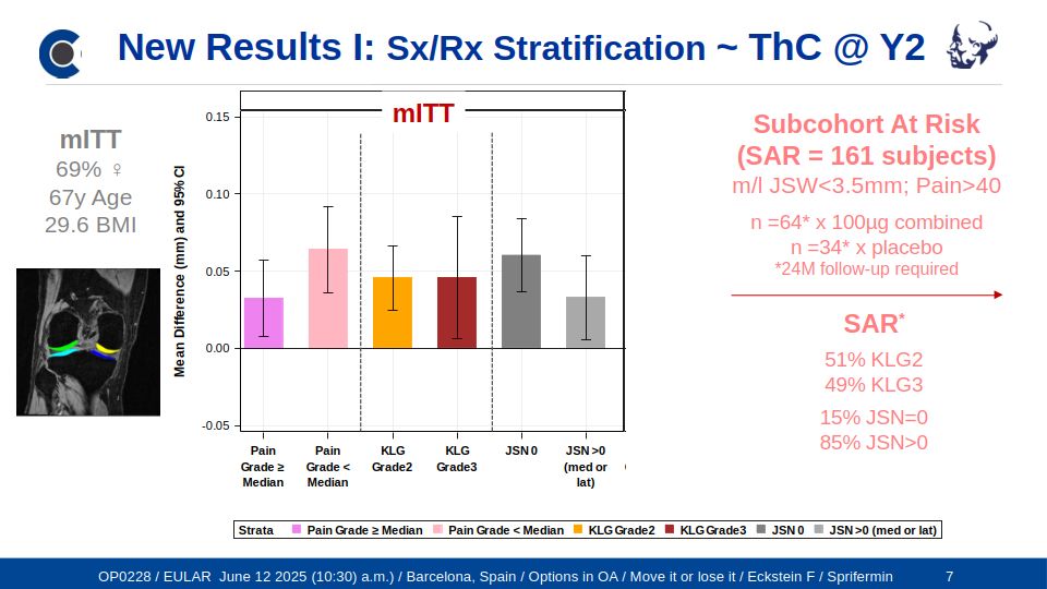
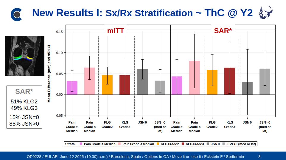
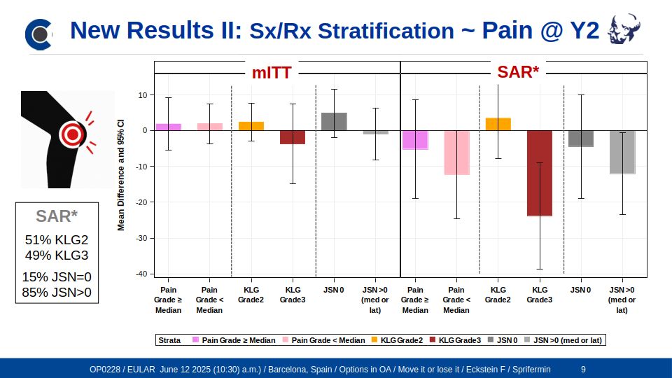
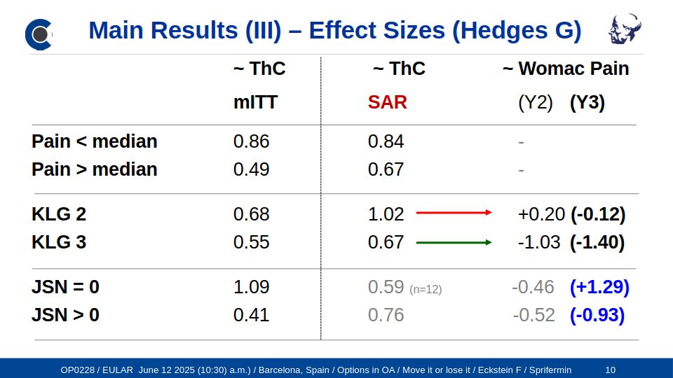
        Pertinent with the inclusion criteria, only a small number of FORWARD participants displayed advanced KOA. In a post-hoc analysis of a subcohort at risk (SAR) (6) that included the patients with more severe radiographic disease and pain, femorotibial cartilage thickness gain with the highest sprifermin dose vs. placebo was as high as for the total cohort (0.06 and 0.05 mm at 2 and 3y, respectively) (6) . In this cohort, with clinically and structurally advanced disease, sprifermin treatment translated into a statistically significant and clinically relevant benefit with the highest dose vs. placebo (-8.75 on a 0-100 WOMAC pain scale vs. only 0.97 for the total cohort) (6) . These findings suggest that the anabolic effect on cartilage is not less pronounced in advanced vs. early KOA, whereas translation of structure modification to symptom benefit appears to be more likely when KOA has progressed further.  

        The 5-year follow-up in FORWARD confirmed that post-treatment cartilage loss was similar amongst the sprifermin- and placebo-treated participants, and that the structural benefit achieved during the treatment period (0.05 mm total femorotibial cartilage thickness between the highest sprifermin dose group vs. placebo) was maintained for another 3 years (7). These findings suggest that the cartilage produced with induction of sprifermin appears to withstand in a normal mechanical environment and provide a lasting benefit after cessation of treatment (7). After 5 years, this structural benefit was also maintained in the SAR, with translation of structure modification into a clinical benefit still apparent after year 5 (7).

        More studies pending publication, also see News Section for recent conference contributions

        1. Dahlberg LE, Aydemir A, Muurahainen N, Gühring H, Fredberg Edebo H, Krarup-Jensen N, et al.
        A first-in-human, double-blind, randomised, placebo-controlled, dose ascending study of intra-articular rhFGF18 (sprifermin) in patients with advanced knee osteoarthritis.
        Clin Exp Rheumatol 2016;34:445–50.

        2. Lohmander LS, Hellot S, Dreher D, Krantz EFW, Kruger DS, Guermazi A, et al.
        Intraarticular sprifermin (recombinant human fibroblast growth factor 18) in knee osteoarthritis: a randomized, double-blind, placebo-controlled trial.
        Arthritis Rheumatol (Hoboken, NJ) 2013;66:1820–1831.

        3. Eckstein F, Wirth W, Guermazi A, Maschek S, Aydemir A.
        Intra-articular sprifermin not only increases cartilage thickness, but also reduces cartilage loss - location-independent post hoc analysis using MR imaging.
        Arthritis Rheumatol 2015;67:2916–2922.

        4. Hochberg MC, Guermazi A, Guehring H, Aydemir A, Wax S, Fleuranceau-Morel P, et al.
        Effect of Intra-Articular Sprifermin vs Placebo on Femorotibial Joint Cartilage Thickness in Patients With Osteoarthritis.
        JAMA 2019;322:1360–1370.

        5. Eckstein F, Wax S, Aydemir A, Wirth W, Maschek S, Hochberg M.
        Intra-articular sprifermin reduces cartilage loss in addition to increasing cartilage gain independent of femorotibial location: a post-hoc analysis of a randomized, placebo-controlled phase ii clinical trial.
        Ann Rheum Dis 2020;79:525–528.

        6. Guehring H, Moreau F, Daelken B, Ladel C, Guenther O, Bihlet AR, et al.
        The effects of sprifermin on symptoms and structure in a subgroup at risk of progression in the FORWARD knee osteoarthritis trial.
        Semin Arthritis Rheum 2021;51:450–456.

        7. Eckstein F, Hochberg MC, Guehring H, Moreau F, Ona V, Bihlet AR, et al.
        Long-term structural and symptomatic effects of intra-articular sprifermin in patients with knee osteoarthritis: 5-year results from the FORWARD study.
        Ann Rheum Dis 2021;80:1062–1069.

        Figure: No of citations of the 20 most cited original articles by Chondrometrics authors

        Figure: No of citations of the 10 most cited review articles by Chondrometrics authors


        Academic Grants, Subcontracts & Services, Software & Training, and Data Sharing Agreements for Medical Image Analysis by Chondrometrics GmbH since its Foundation in 2003.
        More recent contracts are listed on the top; industry contracts are not included.
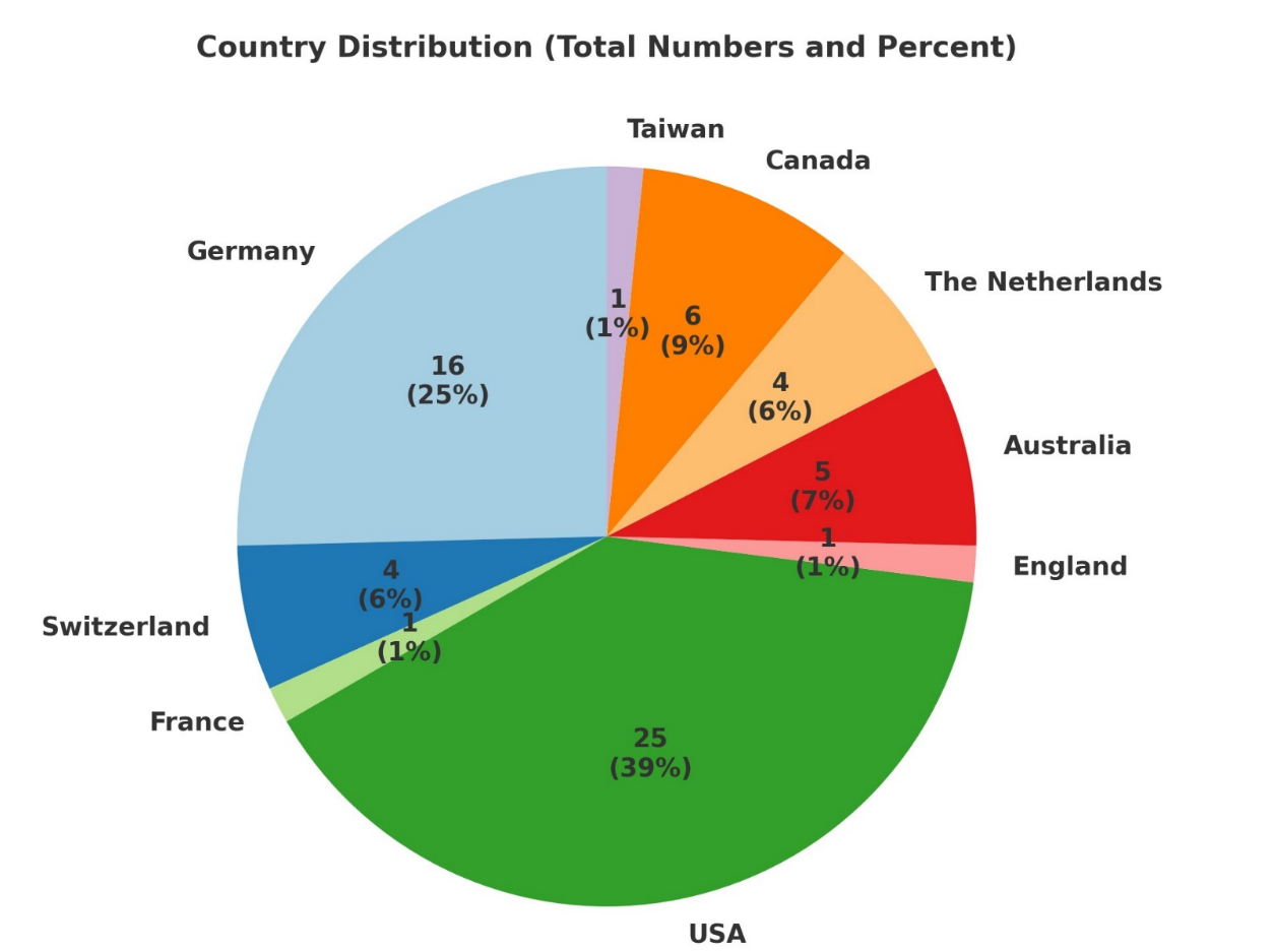
        The pie chart below displays the number of projects per country (percent of total in brackets) from where funding was obtained (63 projects in total).
        Pie Chart Showing the Number of Academic Projects since 2003 per Country (n=63)


        Grant Country Distribution
        • ESA AO-2019-ISS_PP_058 Grant renewed, European Space Agency, through the German Ministry of Economic Affairs and Climate Action (BMWK), Berlin, Germany (Cartilage Degeneration Project); 2023 - 2026
          Felix Eckstein, co-investigator with Anna-Maria Liphardt, Universitätsklinikum Erlangen und Friedrich-Alexander-Universität Erlangen-Nürnberg (#50WB2021), and with Anja Niehoff, Deutsche Sporthochschule Köln (#50WB2022), principal investigators of the project.
          Pre- and post-flight experiments using the International Space Station (ISS) environment: Joint health during a one-year mission to the ISS - an assessment of relevance for exploration
        • ESA AO-20-BR_009 Grant renewed, European Space Agency, through the German Ministry of Economic Affairs and Climate Action (BMWK), Berlin, Germany (Joint Health Project); 2023 - 2026
          Felix Eckstein, co-investigator with Anja Niehoff, Deutsche Sporthochschule Köln (#50WB2022), and Anna-Maria Liphardt, Universitätsklinikum Erlangen und Friedrich-Alexander-Universität Erlangen-Nürnberg (#50WB2021), principal investigators of the project.
          Opportunity soliciting human research proposals for bedrest studies (BRACE und BRAVE)
        • SNF 320030/184912 Grant, University of Basel, Switzerland; 2022-2024
          Felix Eckstein, provider of medical image analysis service, and Annegret Mündermann, University of Basel, principal investigator of the study.
          Analysis of knee cartilage in younger and older participants with or without previous unilateral ACL injury 
        • #E! 114932 EU Grant, Eurostars-2 joint program, with 4P Pharma, Lille, France; 2021-2024
          Co-funding from the European Union Horizon 2020 research and innovation program, with the project management agency (Deutsches Zentrum für Luft- und Raumfahrt) acting on behalf of the Federal Ministry of Education and Research (BMBF). Felix Eckstein & Wolfgang Wirth, work package leads, and Francis Berenbaum, 4P Pharma, principal investigator of the project.
          Lebensverändernde Behandlung für Osteoarthrose-PatientInnen: Ein Biomarker-basierter Lösungsansatz: Teilprojekt: Entwicklung innovativer Bildgebungs-Biomarker für klinische Osteoarthrose-Studien (OA BIO Consortium study)
        • DU PO 1001473784, University of Colorado, Denver, CO, USA; 2021-2024 Felix Eckstein, provider of medical image analysis service, and Jason Dragoo, University of Denver.
          Image analysis with Chondrometrics software in a stem cell chondral defect study and in an osteoarthritis stem cell trial
        • ICL PO, Imperial College, London, England; 2021-2023
          Felix Eckstein and Wolfgang Wirth, provider of medical image analysis service, and Neal Bangerter, Imperial College London, principal investigator of the project.
          Sample cartilage segmentations at 7 Tesla for generating deep learning models facilitating automated cartilage analysis.
        • NHMRC Grant APP1162874, University of Sydney, Australia; 2020-2025.
          Felix Eckstein, provider of clinical trial service, and David Hunter, University of Sydney, principal investigator of the study.
          Stem Cell injections for symptomatic relief and strUctural improvement in people with Tibiofemoral knee OsteoaRthritis (The SCUlpTOR trial))
        • RUG PO, Stichting Ortho Research Center, University Groningen, The Netherlands; 2020-2022
          Wolfgang Wirth & Felix Eckstein, provider of medical image analysis service, and Astrid de Vries, coordinator of the project.
          Use of orthoses in knee osteoarthritis
        • ESA AO-2019-ISS_PP_058 Grant, European Space Agency, through the German Ministry of Economic Affairs and Climate Action (BMWK), Berlin, Germany (Cartilage Degeneration Project); 2019 - 2023
          Felix Eckstein, co-investigator with Anna-Maria Liphardt, Universitätsklinikum Erlangen und Friedrich-Alexander-Universität Erlangen-Nürnberg (#50WB2021), and with Anja Niehoff, Deutsche Sporthochschule Köln (#50WB2022), principal investigators of the project.
          Pre- and post-flight experiments making use of the International Space Station (ISS) environment
        • ESA AO-20-BR_009 Grant, European Space Agency, through the German Ministry of Economic Affairs and Climate Action (BMWK), Berlin, Germany (Joint Health Project); 2019 – 2023
          Felix Eckstein, co-investigator with Anja Niehoff, Deutsche Sporthochschule Köln (#50WB2022), and Anna-Maria Liphardt, Universitätsklinikum Erlangen und Friedrich- Alexander-Universität Erlangen-Nürnberg (#50WB2021), principal investigators of the project.
          Opportunity soliciting human research proposals for bedrest studies (BRACE und BRAVE)
        • FNIH Goods & Service Agreement, Foundation for the National Institutes of Health, Bethesda, MD, USA; 2019-2021
          Felix Eckstein, principal investigator of the service agreement, and Virginia Kraus, Duke University, Durham, NC, USA & David Hunter, University of Sydney, Sydney, NSW, Australia, principal investigators of the project.
          Imaging biomarker analysis service for the Progress OA study (FNIH2)
        • ZonMW Grant 95110008, The Netherlands Organization for Health Research and Development, University Medical Center Utrecht, The Netherlands; 2019-2020
          Felix Eckstein, subcontractor, and Floris Lafeber, University Medical Center Utrecht, principal investigator of the project.
          Joint distraction in treatment of end-stage knee osteoarthritis – Cartilage morphometry in two randomized controlled trials of patients with high tibial osteotomy (HTO) and knee joint distraction (KJD). 
        • SU Data Sharing Agreement between Chondrometrics GmbH and Stanford University, CA, USA; 2019-2020
          Felix Eckstein and Akshay Chaudhari, Department of Radiology, Standford University, Co-PI of the project.
          A machine- (deep-) learning approach to fully automated regional bone, cartilage and phenotype analysis in knees osteoarthritis – development, validation, application, and qualification
        • DSHS PO 4500001636, Deutsche Sporthochschule Köln, Germany; 2019-2020
          Felix Eckstein, provider of medical image analysis software and training, and Peter Brüggemann, Deutsche Sporthochschule Köln, principal investigator of the study.
          Measurement of cartilage properties in relation to the use of footwear
        • UWO PO 181229, University of Western Ontario, London, Canada; 2018-2024
          Felix Eckstein, provider of medical image analysis service (articular cartilage morphology and transverse relaxation time), and Trevor Birmingham, University of Western Ontario, principal investigator of the project.
          Longitudinal analysis of cartilage MRI properties before and after high tibial osteotomy (HTO).
        • EU IMI2 Grant 115770 (Subcontract), European Union Innovative Medicines Initiative (IMI) by University Medical Center Utrecht, The Netherlands; 2018-2021
          Felix Eckstein, principal investigator of the subcontract, and Harry Weinans, University Medical Center Utrecht, principal investigator of the work package.
          Applied public-private research enabling osteoarthritis clinical headway (APPROACH study).
        • PO DFKI IIP-EXTREM, Deutsches Forschungszentrum für Künstliche Intelligenz, Kaiserslautern, Germany; 2018-2020
          Felix Eckstein, provider of medical image analysis service, and Christian Schlinkmann, Deutsches Forschungszentrum für Künstliche Intelligenz.
          Segmentation of anatomical structures of the thigh and shank from magnetic resonance images (MRIs) for the generation of automated models for generating personalized prostheses.
        • SU PO 61913492, Stanford University, Redwood City, CA, USA; 2018-2020 Felix Eckstein, provider of medical image analysis service, and Jason Dragoo, Department of Orthopaedic Surgery, Stanford University.
          Quantitative analysis of femoral cartilage in a stem cell chondral defect study and in an osteoarthritis stem cell trial
        • Investigator Initiated Co-Funding Project (IIP) between Chondrometrics GmbH and Merck KGaA, Germany; 2018-2019
          Felix Eckstein, PI of the IIP, and Christoph Ladel, Merck KGaA, Co-PI of the project.
          Determination of thresholds of progression from the healthy reference cohort of the Osteoarthritis Initiative (OAI) with coronal FLASH and sagittal DESS imaging
        • NTUST PO, National University of Science and Technology, Taipeh, Taiwan; 2018-2019
          Felix Eckstein, provider of medical image analysis service, and Wei-Chun Hsu, Graduate Institute of Biomedical Engineering Taipei, principal investigator of the project.
          Study of an athletic population with unilateral symptomatic osteoarthritis. 
        • DFG Grant NE 2136/3-1, 701010997 Deutsche Forschungsgemeinschaft, Universitätsklinikum Düsseldorf, Germany; 2018-2019
          Felix Eckstein, provider of medical image analysis service, and Sven Nebelung, University Düsseldorf, principal investigator of the project.
          Functional and biomechanical methods using multi-parametric MRI for early detection of osteoarthritis. 
        • SU PO 61763686, Stanford University, Palo Alto, CA, USA; 2018-2019
          Felix Eckstein, provider of medical image analysis service, and Constance Chu, Stanford University, principal investigator of the study.
          Femoral cartilage transverse relaxation time (T2) in an academic trial on effects of unloader bracing on clinical outcome and articular cartilage regeneration following microfracture of isolated chondral defects. 
        • Investigator Initiated Co-Funding Project (IIP) between Chondrometrics GmbH and Merck KGaA, Germany; 2017-2018
          Felix Eckstein, PI of the IIP, and Christoph Ladel, Merck KGaA, Co-PI of the project.
          Prediction of incident radiographic osteoarthritis by 3D meniscus morphology and position properties from MRI, in knees with early osteoarthritis (KLG1)
        • SickKids PO REB FN10000010841, Hospital for Sick Children, Toronto, Canada; 2016-2017
          Felix Eckstein, provider of medical image analysis service, and Andrea Doria, Hospital for Sick Children, principal investigator of the study.
          Analysis of ankle cartilage in patients with hemophilia and controls
        • Investigator Initiated Co-Funding Project (IIP) between Chondrometrics GmbH and Merck KGaA, Germany; 2016-2017
          Felix Eckstein, PI of the IIP, and Martin Michaelis, Merck KGaA, Co-PIs of the project.
          Prediction of incident radiographic osteoarthritis by cartilage composition (MRI transverse relaxation time) in knees with early osteoarthritis (KLG1)
        • ESA Res. Announcement ILSRA-2009-BR-0994 & German Federal Ministry for Economic Affairs (BWi) 50WB1520 Grant, Berlin, Germany; 2015-2019.
          Felix Eckstein, co-investigator with Anja Niehoff, Deutsche Sporthochschule Köln, principal investigator of the project.
          The effects of microgravity on cartilage morphology and biology
        • UTAS Research Enhancement Grant, University of Tasmania, Hobart, Australia; 2015-2016 Felix Eckstein, provider of medical image analysis service; and Benny Eathakkattu Antony, University of Tasmania, principal investigator of the grant.
          Associations of physical activity, physical performance measures and obesity in childhood with knee cartilage thickness in adults after 25 years
        • Canadian Institute of Health Research (CIHR) Grant, University of Western Ontario, London, Canada; 2014-2019.
          Felix Eckstein, provider of medical image analysis software and training, and Trevor Birmingham, University of Western Ontario, principal investigator of the project.
          Load-altering interventions and knee osteoarthritis progression.
        • SickKids PO REB FN 10000010841, Hospital for Sick Children, Toronto, Canada; 2014-2015
          Felix Eckstein, provider of medical image analysis service, and Andrea Doria, Hospital for Sick Children, principal investigator of the study.
          Measurement properties of ultrasound for assessment of hemophilic arthropathy: MRI correlation
        • DHS PO 13-20, Deutsche Sporthochschule, Köln, Germany; 2013-2015
          Felix Eckstein, provider of medical image analysis software and training, and Anja Niehoff, Deutsche Sporthochschule Köln, principal investigator of the study.
          Measurement of knee cartilage morphology in biomechanically relevant studies
        • Investigator Initiated Co-Funding Project (IIP) between Chondrometrics GmbH and Merck KGaA, Germany; 2013-2014
          Felix Eckstein, PI of the IIP, and Christoph Ladel, Merck KGaA, Co-PI of the project.
          Predictive value of imaging biomarkers (cartilage morphology at T-3 and T-1) on risk of total knee replacement at 48-60 months in the Osteoarthritis Initiative (OAI)
        • NIH P60 Grant AR047785, Multidisciplinary clinical research center (MCRC); National Institutes of Health, Bethesda, USA; 2012-2017 Felix Eckstein, principal investigator of subcontract; David Felson, Boston University, Boston, MA, USA, principal investigator of the grant.
          Effect of bariatric surgery on knee structure and pain (OABS study)
        • FNIH Goods & Service Agreement, Foundation for the National Institutes of Health, Bethesda, MD, USA; 2012-2014
          Felix Eckstein, principal investigator of the service agreement, and Virginia Kraus, Duke University, Durham, NC, USA & David Hunter, University of Sydney, Sydney, NSW, Australia, principal investigators of the project.
          Imaging biomarker analysis service for the Osteoarthritis Initiative (OAI) Biomarker Consortium Study (FNIH1)
        • Investigator Initiated Co-Funding Project (IIP) between Chondrometrics GmbH and Merck KGaA, Germany; 2012-2013
          Felix Eckstein, PI of the IIP, and Christoph Ladel, Merck KGaA, Co-PI of the project.
          Longitudinal analysis of cartilage morphology in the in the OAI healthy reference cohort and in the Foundation of NIH OAI Biomarker Consortium Sample (cont.).
        • RN LLP-9 Grant, ReumaNederland (Dutch Arthritis Society), The Netherlands; 2011-2016
          Felix Eckstein, subcontractor, and Floris Lafeber, University Medical Center Utrecht, principal investigator of the project.
          Mechanisms of tissue degeneration and regeneration in rheumatic diseases with focus on joint distraction therapy in osteoarthritis. 
        • FHS USYD PO, Faculty of Healthy Sciences, University of Sydney; Australia 2011-2012
          Felix Eckstein, provider of medical image analysis software and training, and Marlene Fransen, University of Sydney, principal investigator of the study.
          Longitudinal measurement of knee cartilage morphometry using MR imaging, to evaluate the benefit of glucosamine sulfate, with and without chondroitin sulfate, for people with symptomatic knee osteoarthritis in terms of structural knee progression.
        • NIH NHLBI Grant HHSN26820100002, National Institutes of Health & National Heart, Lung, and Blood Institute, Bethesda, MD, USA; 2010-2015 Felix Eckstein, principal investigator of the subcontract, Kent Kwoh, University of Pittsburgh, PA, USA, principal investigator of the project.
          Pivotal Osteoarthritis Initiative MRI Analyses (the POMA study)
        • Investigator Initiated Co-Funding Project (IIP) between Chondrometrics GmbH and Novartis Pharma AG, Basel, Switzerland; 2010-2011
          Felix Eckstein, PI of the IIP, and Markus John, Novartis Pharma AG, Co-PI of the project.
          Prognostic value of clinical, serological and imaging biomarkers on risk of total knee replacement (TKR): a prospective observational cohort study in patients with knee osteoarthritis.
        • Investigator Initiated Co-Funding Project (IIP) between Chondrometrics GmbH and Merck KGaA, Germany; 2010-2011
          Felix Eckstein, PI of the IIP, and Christoph Ladel, Merck KGaA, Co-PI of the project.
          Longterm (4-year) longitudinal analysis of cartilage morphology change in the Foundation of NIH (FNIH) OAI Biomarker Consortium Sample
        • NIH R01 Grant AR054806, National Institutes of Health, Bethesda, MD, USA; 2009-2013 Felix Eckstein, principal investigator of the subcontract, and Leena Sharma, Northwestern University, Chicago, MI, USA, principal investigator of the project.
          Relationship of hip muscle group weakness to deterioration of the OA knee by MRI
        • Investigator Initiated Project (IIP OAI No8) between Chondrometrics GmbH and Novartis Pharma AG, Basel, Switzerland, 2008-2009
          Felix Eckstein, PI of the IIP, and Alberto Gimona, Novartis Pharma AG, Co-PI of the project.
          Advanced radiographic disease (KLG 4) two- (and one-) year longitudinal analysis of cartilage morphology change in a sample of the Osteoarthritis Initiative 
        • Investigator Initiated Project (IIP OAI No7) between Chondrometrics GmbH and Centocor Inc. R&D, USA; 2008-2009
          Felix Eckstein, PI of the IIP, and Frederic Baribaud, Centocor R&D, Co-PI of the project.
          Healthy reference cohort analysis of longitudinal change in cartilage morphology in the Osteoarthritis Initiative
        • Investigator Initiated Project (IIP OAI No6) between Chondrometrics GmbH and Wyeth Co, USA; 2008-2009
          Felix Eckstein, PI of the IIP, and Jennifer Lee, Wyeth, Co-PI of the project.
          Subregion analysis of two- (and one-) year longitudinal change in cartilage morphology in the Osteoarthritis Initiative.
        • Investigator Initiated Project (IIP OAI No5) between Chondrometrics GmbH and the Osteoarthritis initiative (OAI) Coordinating Center (UCSF Vendor Contract 9000011523), University of California, San Francisco, CA, USA; 2008-2009
          Felix Eckstein and Michael Nevitt, University of California San Francisco, co-principal investigators of the project.
          Two- (and one-) year longitudinal analysis of cartilage morphology in a further sample of the Osteoarthritis Initiative using sagittal double echo steady state MRI
        • Investigator Initiated Project (IIP OAI No4) between Chondrometrics GmbH and GlaxoSmithKline (“GSK”), USA; 2008-2009
          Felix Eckstein, PI of the IIP, and Dominique Ethgen, GlaxoSmithKline, Co-PI of the project.
          Two-year longitudinal analysis of cartilage morphology in a further sample of the Osteoarthritis Initiative using coronal spoiled gradient echo MRI.
        • Investigator Initiated Project (IIP OAI No3) between Chondrometrics GmbH and Merck Serono, Switzerland; 2008-2009
          Felix Eckstein, PI of the IIP, and Alberto Gimona, Merck Serono, Co-PI of the project.
          One-year longitudinal analysis of cartilage morphology change in a further sample of the Osteoarthritis Initiative, using coronal spoiled gradient echo MRI.
        • Investigator Initiated Project (IIP OAI No2) between Chondrometrics GmbH and Eli Lilly & Co, USA; 2007-2008
          Felix Eckstein, PI of the IIP, and Olivier Benichou, Eli Lilly & Co, Co-PI of the project.
          One-year longitudinal analysis of side-differences in cartilage morphology change in subjects from the Osteoarthritis Initiative with uni-lateral radiographic joint space narrowing, using from sagittal dual echo steady state (DESS) MRI.
        • Investigator Initiated Project (IIP OAI No1) between Chondrometrics GmbH and Pfizer Inc., USA; 2006-2008
          Felix Eckstein, PI of the IIP, and Marie-Pierre Hellio Le Graverand, Pfizer Inc., Co-PI of the project.
          One-year longitudinal analysis of cartilage morphology change in a first progressor sample of the Osteoarthritis Initiative using coronal spoiled gradient echo MRI.
        • NIH P60 MCRC Grant AR048098, National Institutes of Health, Bethesda, MD, USA; 2006-2012
          Felix Eckstein, principal investigator of the subcontract, Leena Sharma, Northwestern University, Chicago, MI, USA, principal investigator of the project & Richard Pope, Northwestern University, principal investigator of the Multidisciplinary Clinical Research Center.
          Potential Beneficial Role of Hip Muscles in Knee Osteoarthritis.
        • UCSF Vendor Contract 9000011523, Osteoarthritis Initiatives (OAI) Coordinating Center, University of California,, San Francisco, CA, USA; 2006-2008 Felix Eckstein, principal investigator of the subcontract, with Michael Nevitt, University of California, San Francisco, principal investigator of the project.
          Baseline, 12, and 24 month quantitative cartilage analysis in a sample of Osteoarthritis Initiative index knees (OAI)
        • NIH RO1 Grant AR052528-01A2 National Institutes of Health, Bethesda, MD, USA; 2006-2011
          Felix Eckstein, principal investigator of the subcontract, and Stephen Messier, Wake Forest University, Winston Salem, NC, USA, principal investigator of the project.
          Intensive dietary restriction with exercise in arthritis (IDEA trial)
        • ACRIN Grant, American College of Radiology, USA; 2006-2008:
          Felix Eckstein, principal investigator of the sucontract, with Timothy Mosher, Pennsylvania State University, PA, USA, principal investigator of the project.
          Reproducibility & stability of cartilage morphology measures with 3 Tesla MRI.
        • RNSH PO, Institute of Bone and Joint Research, Royal North Shore Hospital, Sydney, Australia; 2006-2008.
          Felix Eckstein, provider of medical image analysis software and training, and Lyn March, Royal North Shore Hospital, principal investigator of the study.
          Measurement of cartilage morphology for assessing the effect of weight loss on knee joint structure in patients undergoing two different weight loss programs.
        • USYD PO, School of Exercise and Sport Science, University of Sydney, Australia; 2006-2008.
          Felix Eckstein, provider of medical image analysis software and training, and Benedicte Vanwanseele, School of Exercise and Sport Science; principal investigator of the study.
          Relationship of cartilage/meniscus morphology with gait biomechanics parameters in women with knee osteoarthritis
        • NIH R01 Grant AR52918, National Institutes of Health, Bethesda, MD, USA; 2005-2010
          Felix Eckstein, principal investigator of the subcontract, and Leena Sharma, Northwestern University, Chicago, MI, USA, principal investigator of the project.
          Ancillary study to the Osteoarthritis Initiative (OAI)
        • DSHS PO 100202, Deutsche Sporthochschule, Köln, Germany; 2005-2007
          Felix Eckstein, provider of medical image analysis software and training, and Peter Brüggemann, Sporthochschule Köln, principal investigator of the study.
          Knee joint cartilage morphology in athletes before, and after specific impact exercise protocols, and comparison with physically inactive volunteers
        • McM PO, McMaster University, Hamilton, Canada; 2005-2007
          Felix Eckstein, provider of medical image analysis software and training, and Karen Beattie, Department of Medicine, McMaster University, Hamilton, ON, Canada; investigator of the study.
          Comparison of quantitative cartilage parameters in healthy and arthritic knees and comparison with minimum joint space width on fixed flexion radiographs. from MR images in paraplegic patients versus controls
        • NIH RO1 Grant HD043500, National Institutes of Health, Bethesda, MD, USA; 2005-2007
          Felix Eckstein, principal investigator of the subcontract, and Leena Sharma, Northwestern University, Chicago, MI, USA, principal investigator of the project.
          Laxity and malalignment in a large cohort study of OA (MOST study)
        • CFI (Canada Foundation for Innovation) infrastructure grant; University of British Columbia, Vancouver; 2004-2007.
          Felix Eckstein, provider of medical image analysis software and training, and David Wilson, University of British Columbia, principal investigator of the study. Measurement of cartilage morphometric properties from MR images of the human hip and shoulder
        • NIH funded UCSF Vendor Contract, Osteoarthritis Initiatives (OAI) Coordinating Center, University of California, San Francisco, CA, USA, by the National Institutes of Health, Bethesda, MD, USA; 2004-2005
          Felix Eckstein, principal investigator of the subcontract, with Michael Nevitt, University of California, San Francisco, principal investigator of the project.
          Test-retest cartilage volume analysis for cross validation and coil comparison in the Osteoarthritis Initiative (OAI) pilot study.
        • NIH RO1 Grant AR48216 National Institutes of Health, Bethesda, MD, USA; 2003-2007
          Felix Eckstein, principal investigator of the subcontract, and Leena Sharma, Northwestern University, Chicago, principal investigator of the project.
          Progression of knee OA: The role of local factors (the MAK study).
        • NIH P60 Center Grant AR47785, National Institutes of Health, Bethesda, MD, USA; 2003-2006
          Felix Eckstein, principal investigator of the subcontract, and David Felson, Boston University, Boston, MA, USA, principal investigator of the project.
          Correlates of a rticular cartilage thickness in knees of subjects in the Framingham study.
        • AF Clinical Sciences Grant, Arthritis Foundation, Atlanta, GA, USA; 2003-2006
          Felix Eckstein, principal investigator of the subcontract, and David Felson, Boston University, Boston, MA, USA, principal investigator of the project.
          Correlates of articular cartilage volume and thickness in Framingham subjects.

        SAB & PARTNERS

        Scientific Advisory Board (SAB)

        PMU Partner Neal K. Bangerter
        Neal Bangerter

        Neal Bangerter is an Associate Professor of Bioengineering at Imperial College London. His current research interests include the application of artificial intelligence and machine learning to a variety of problems in medical imaging and healthcare, and the development of novel medical imaging technologies with a particular focus on musculoskeletal applications.

        In addition to his research and teaching interests, Neal is passionate about innovation/entrepreneurship and intellectual property strategy. He regularly consults and advises in these areas. He has deep technical expertise in healthcare, medical imaging, machine learning, big data, signal processing, and software development, and significant management and strategy experience from his work in industry.

        Neal holds a BA with honors in Physics from UC-Berkeley and a Master’s and PhD in Electrical Engineering from Stanford University. He has held permanent and visiting positions at Oxford University, INSEAD, Brigham Young University, University of Utah, Microsoft, and management consulting firm McKinsey & Company.

        Mikael Boesen
        CV Boesen
        Name: Mikael Ploug Boesen, MD, Ph.D.
        Born: May 27th 1971 in Copenhagen, Denmark
        Work: Bispebjerg and Frederiksberg Hospital, Deptof Radiology, Bispebjerg Bakke 23, 2400 Copenhagen NV, Denmark

        2017 Professor in musculoskeletal radiology at Copenhagen University and Bispebjerg and Frederiksberg Hospital
        2015 Associate Professor in Radiology at Copenhagen University, Denmark
        2014 Senior Consultant, head of musculoskeletal imaging and research, Dept. of Radiology, Bispebjerg and Frederiksberg Hospital
        2009-2013 Head of MSK MRI research, Dept. of Radiology, Bispebjerg and Frederiksberg
        2013 Specialist in Radiology, Consultant in Radiology, Dept. of Radiology Bispebjerg and Frederiksberg
        2008 PhD degree entitled: “Magnetic Resonance Imaging of Joints Following Intra-articular Treatment and Procedures in Arthritis”
        1999 Medical school graduation (Cand. Med.), University of Copenhagen

        I defended my PhD thesis in June 2008, entitled "Magnetic resonance imaging of joints following intra-articular treatment in arthritis" from the Parker Institute and Aalborg University. Throughout my radiology career I have pursued my special interest in advanced imaging of patients with various musculoskeletal conditions. My group and I have, specialised in quantification of cartilage changes as well as, soft tissue and bone inflammation in the joints with advanced imaging. We also serve as European reference centre for studies using a tilting open dedicated MRI unite – G-scan from ESAOTE, looking at the mechanical changes in the soft tissue, bone and cartilage in the spine, knee joints and feet going from supine to standing position.

        Since 2008 I have been chairman of the IMAGINE group investigating DCE-MRI in patients with inflammatory arthritis, and I have co-organised several national and international imaging symposia’s and imaging courses, and have served as radiological specialist in the development of the national and international guidelines for imaging of degenerative and inflammatory joint and spinal diseases. I am an invited associate board member of the of the Arthritis Research UK Centre for Sport, Exercise and Osteoarthritis, MUSKRAD, ESSR, DRS, ESR, EULAR,OARSI, DIMS and I am part of the clinical and scientific advisory board of Image Analysis Group, London UK since 2010, where I have served as chairman until January 2019. Recently I have joined the scientific advisory board member of Chondrometrics, GmbH, Ainring, Germany in February 2019.

        Management experience
        Jan 2014- Senior Consultant, head of musculoskeletal imaging and research, Dept. of Radiology, Bispebjerg and Frederiksberg Hospital, Copenhagen Denmark
        2010-2018 Head of clinical and research advisory board Image Analysis Group (IAG), London, UK
        2007- Head of MSK MRI research, Dept. of Radiology, and Parker Institute, Bispebjerg and Frederiksberg Hospital, Copenhagen Denmark

        Supervised or co-supervised, 4 pre-graduate students, 15 national PhD’s (6 as main supervisor) and 2 international PhD’s as well as 2 postdocs


        Link to complete list of publications:Research gate profile, Google Scholar
        Rainer Burgkart
        Rainer Burgkart

        Rainer Burgkart was born in Munich, Germany. He studied Medicine in Essen and Münster and graduated in 1988. From 1989 to 1991 Rainer Burgkart was resident in the Clinic for Orthopaedics and Sportorthopaedics at the Klinikum r.d. Isar der Technical University Munich (TUM). From 1991 to 1992 he received a research fellowship in the Orthopaedic Clinic, Duke University/Durham, N.C., USA (Direktor: Prof. Dr. J. Urbaniak). After continuing residency in 1992 in Clinic for Orthopaedics and Sportorthopaedics at the Klinikum r.d. Isar he completed his board exams in Juli 1997. Since then he worked as a senior attending (Oberarzt) at this Clinic and received a second research fellowship and joined the research group of Prof. Farsh Guilak from 1998 to 1999 in the Orthopaedic Clinic, Duke University/Durham, N.C., USA.

        Since 2000 Rainer Burgkart is Head of Research and Education of the Clinic for Orthopaedics and Sportorthopaedics, Klinikum rd Isar, TUM. He completed his “Habilitation” in 2007 and since February 2007 announced as an Associate Professor.

        His research focus are topics as MRT cartilage characterization and Ultrasound Imaging including AI-based analytic methods, Endoprostheses, Biofunctional Implant Surfaces in respect to Bone Ingrowth and Antibacterial Properties (in vitro (cell lab) and in vivo testing (animal), Computer Assisted Surgery and Robotics, Implant Testing and Implant Optimization, Biomechanics of Macro- and Micro- Level (e.g. subcellular Biomechanics in Tissue Culture) as well as Simulators for Medical Education (especially haptic interfaces), new Training Systems for Musculoskelettal Exercise, Virtual Operation Planning, Modelling and AI.

        Rainer Burgkart was responsible Congress Secretary of the German Orthopaedic Society (DGOOC) for organizing the first common German Congress of Orthopaedic and Traumatology (DKOU) in Berlin in 2005. In addition he was involved in the organisation of several congress as the 5th World Congress of Biomechanics in 2006 in Munich and the Congress of the German Surgery Society in Berlin in 2010. He was the founding member of the German “Netzwerk für Regenerative Medizin” and the “Netzwerk für Muskuloskelettale Biomechanik”. From 2006 until 2019 he was a Board member of DFG- Excellenz-Initiative TUM International Graduate School of Science and Engineering (IGSSE) and since 2007 he is a permanent Board member of the „Deutscher DIN Normenausschuss DIN NA 027-02-15 AA - Endoprothetik und Osteosynthese".

        As a selection of scientific awards Rainer Burgkart received e.g. for cartilage analysis the Göran Selvik Award of European Orthopaedic Research Society 1999 (for advanced computer 3D volumetry methods in „MRI-based assessment of cartilage loss in severe osteoarthritis- accuracy, precision and diagnostic value“) and the Investigator Award des V. Münchener Symposium in 2015 (for “Kartographie des Erholungsverhaltens und der Steifigkeit des artikulären Knorpels im adulten, ovinen Kniegelenk) and for roboter-based new teaching simulators e.g. the ISPO Academic Challenge Award 2003, “The Best of the Best“- Award of the Bayerische Gesellschaft für Geburtshilfe und Frauenheilkunde e.V. (BGGF) 2004, the EU Robotics Technology Transfer Award 2011, and in the field of tissue characterization the Winner of the Bionikwettbewerb of TUM 2008 for the project „Hard-Soft Interfaces in Biology and Technical Applications – e.g. the tendon-bone interface“, the Karl-Heinz-Höhne 1. Prize MedVis-Award 2010 for „Innovative Visualizing of Implant Planning in Orthopedics” and as Teaching Award e.g. the Excellence Award of "Best Academic Teacher of 2015 in TUM Medicine".

        Akshay Chaudhari
        CV Chauddhari

        Akshay Chaudhari is a Research Scientist at the Department of Radiology at Stanford University. Akshay graduated with a Ph.D. in Bioengineering from Stanford University in 2017 and B.S. from the University of California San Diego in 2012. He is interested intertwining medical imaging and computer science to develop biomedical imaging methodologies to better under physiology and to enhance the efficacy and efficiency of current diagnostic techniques. Akshay is currently combining deep learning and computer vision methods to advanced magnetic resonance imaging (MRI) data acquisition and signal processing techniques for generating rapid, quantitative and morphological magnetic resonance imaging (MRI) examinations. He seeks to develop such MR imaging techniques and implement them for diagnostic clinical imaging and in research studies evaluating early disease progression.

        Akshay is experienced with hardware and software start-up companies. He previously was a consulting Technology and Application Specialist for Skope Magnetic Resonance Technologies and is currently a Deep Learning Research Scientist at Subtle Medical and also a scientific advisor for Brain Key.


        LinkedIn Profile
        Google Scholar Profile
        Jamie E. Collins 
        CV Collins

        Jamie E. Collins is Associate Professor of Orthopaedic Surgery at Harvard Medical School and Associate Director of the Orthopaedic and Arthritis Center for Outcomes Research at Brigham and Women’s Hospital in Boston, USA. She is the Director of the Biometry Consultancy Unit in the Department of Orthopaedic Surgery at Brigham and Women’s Hospital and Director of the Data Management & Analysis Core of the Brigham Coordinating Center for the Arthritis Foundation’s Osteoarthritis Clinical Trials Network. She received her master’s degree in mathematics and PhD in Biostatistics from Boston University.  

        Her research interests include the analysis of multilevel and correlated data, missing data and informative dropout, group-based trajectory and latent growth curve models, risk prediction, and machine learning methods for clustering and disease phenotyping. She oversees data collection and analysis for a number of NIH and foundation funded randomized controlled trials in OA and rheumatology.

        Jamie is the lead statistician for the FNIH OA Biomarkers Consortium, an international collaboration that aims to identify novel knee osteoarthritis biomarkers with the greatest utility for predicting disease progression and treatment response in clinical trials. This public-private partnership is supported by the NIH, FDA, Arthritis Foundation, and a number of industry partners. The ultimate goal is formal biomarker qualification by regulatory authorities such as FDA and EMA.

        Jamie is an Associate Editor for Statistics at Osteoarthritis and Cartilage, an Advisory Editor for Statistics at Arthritis & Rheumatology, and an Editorial Board Member at Arthritis Care and Research and Osteoarthritis Imaging. She is chair of the Osteoarthritis Research Society International Communications Committee and a member of the American College of Rheumatology Committee on Registries and Health Information Technology. 

        Chris Ladel
        CV Chris Ladel

        Chris Ladel is a seasoned, forward-thinking, and highly skilled project manager and consultant with broad experience in several areas of pharmaceutical development. He was originally trained as an immunologist in preclinical and clinical investigations in different disease areas including infectious diseases, autoimmune diseases, and osteoarthritis. Following post-doctoral fellowship, he gained several years of industry experience with pharmaceutical companies such as GlaxoWellcome/GSK, Bayer, MerckSerono/Merck KGaA, where he was primarily involved in leading teams on strategy generation and in-licensing/due-diligence activities; he also represented the companies in national and international consortia, and outsourced activities for validation and qualification of biomarkers.

        The clinical research experience focus over the last two decades has been on the development of therapies in immunology, rheumatology, and osteoarthritis, with particular interest in personalized medicine and stratification approaches in clinical development, using biomarkers and imaging techniques. He has been involved in several patents and publications on this topic and continue to seek out opportunities that require development of clinical strategies in these areas.

        Chris is also skilled in collaboration and project management. At Merck, he was responsible for international collaborations in non-clinical as well as clinical settings via different public-private initiatives with a focus on qualification and validation of biomarkers. Additionally, Chris has served as project lead in several endeavors from early research up to phase II clinical trials with a history of successful clinical development. As a core team member of several programs with focus on translational aspects and clinical biomarker strategies, he also has been involved in operational activities, including budget planning for programs and portfolios in defined therapeutic areas. Further, Chris is experienced in consultancy, as he has provided consultant services for several companies at early clinical stages to develop strategies, including translational approaches for different entities.


        Linkedin Profile
        Research Gate
        Ali Mobasheri
        CV Chris Mobasheri

        Ali Mobasheri is Professor of Musculoskeletal Biology in the Research Unit of Health Sciences and Technology within the Faculty of Medicine at the University of Oulu in Finland. He also holds the position of Chief Researcher and International Adviser in the State Research Institute Centre for Innovative Medicine in Vilnius, Lithuania. He is the Immediate Past President of the Osteoarthritis Research Society International (OARSI) and serves on the Scientific Advisory Board of the European Society for Clinical and Economic Aspects of Osteoporosis, Osteoarthritis and Musculoskeletal Diseases (ESCEO). In addition, he serves as “Collaborateur Scientifique de l’Université de Liège” and Senior Strategic Advisor to the World Health Organization Collaborating Center for Public Health Aspects of Musculoskeletal Health and Aging, Université de Liège, Liège, Belgium. He is a member of the International Cartilage Regeneration & Joint Preservation Society (ICRS) and serves on the translational science committee of ICRS.

        Ali has held visiting professorships at Harvard University in Boston (USA), University Medical Centre Utrecht (Netherlands) and King Abdulaziz University in Jeddah (Kingdom of Saudi Arabia). He is currently a Visiting Professor at Sun Yat-sen University, Guangzhou, Guangdong, China. His current research focuses on cartilage biology, specifically cellular metabolism, ion channel physiology and biomarkers of osteoarthritis. He is ranked as one of the top 10 leading experts in the world on “osteoarthritis” and “cartilage” on expertscape.com. Ali already leads a multidisciplinary team of researchers in Vilnius and is building a new team of researchers at the University of Oulu. His work is currently funded by the Academy of Finland, The Lithuanian Research Council, and several funding mechanisms of the European Commission (FP7, Horizon Europe, COST Action, EuroNanoMed, Innovative Medicines Initiative).


        Current Positions:
        • Professor of Musculoskeletal Biology in the Fibrobesity Research Theme and the Research Unit of Health Sciences and Technology, Faculty of Medicine, University of Oulu, Aapistie 5 A, FIN-90230 Oulu, Finland
        • Chief Researcher and International Advisor, State Research Institute C entre for Innovative Medicine, Santariskiu 5, 08661 Vilnius, Republic of Lithuania
        • Immediate Past President, Osteoarthritis Research Society International (OARSI) and Member of the OARSI Board of Directors
        • Visiting Professor, Sun Yat-sen University, Guangzhou, Guangdong, China

        Recent Visiting Positions:
        • Associate Professor, Departments of Orthopaedics, Rheumatology and Clinical Immunology, University Medical Centre Utrecht, Utrecht, The Netherlands (March 2020 to October 2021)
        • Visiting Professor, Harvard University, Boston, USA (Non-Stipendiary position) (March 2020 to March 2021)
        • Distinguished Adjunct Professor (Thomson ISI Highly Cited Program), King Abdulaziz University, Jeddah, Kingdom of Saudi Arabia (February 2013 to May 2016)
        Tom Turmezei
        CV Tom Turmezei

        Tom Turmezei has been a Consultant Radiologist at the Norfolk and Norwich University Hospital, UK, since 2017 and has over a decade of specialist diagnostic and interventional experience in a wide range of musculoskeletal disorders, including joint disease. Having graduated from the University of Cambridge, UK, in 1999 (MA) and then Oxford, UK, in 2002 (BMBCh), he trained in Radiology at Norwich, Nottingham and Cambridge, followed by a musculoskeletal specialist fellowship at the Royal National Orthopaedic Hospital in Stanmore, UK, from 2016 to 2017.

        Promoted to Honorary Professor at the University of East Anglia, UK, in 2022, Tom spent three years between 2012 and 2016 in his Wellcome Trust clinical PhD fellowship with the Medical Imaging Group at the Cambridge University Engineering Department, UK. During this time he developed quantitative 3-D computed tomography (CT) and magnetic resonance imaging (MRI) analysis techniques for joint space and articular cartilage evaluation. Having evolved these throughout the lower limb, his research has also included cartilage segmentation and multiparametric MRI at the knee joint.

        Tom has twice received the Young Investigator Award from the International Workshop of Osteoarthritis Imaging (2016 & 2019), and is organising the 2025 meeting in Cambridge, UK, where he is also currently a visiting researcher in the University Departments of Medicine and Orthopaedic Surgery.

        Tom has worked in a number of international collaborations, including with the Multicenter Osteoarthritis Study (USA) and the AGES-Reykjavik cohort (Iceland), and has supervised postgraduate students at the University Medical Center Utrecht (Netherlands), the University of Oulu (Finland), and the University of Copenhagen (Denmark).

        Tom is a Scientific Advisory Board member for the International Society of Osteoarthritis Imaging and founding chair of the Computed Tomography in Osteoarthritis (OCTA) Research group. With clinical and research experience in both osteoarthritis and osteoporosis, he has provided consultancy services to Curvebeam AI, Pfizer Inc., GSK plc, and Amgen Inc., and has oversight of imaging for more than 150 clinical trials in his current position as Imaging Research Lead in Norwich.

        Tom is also an award-winning writer and editor of academic textbooks and is currently the Imaging Editor for Gray’s Anatomy (Elsevier). He is a long-standing advocate for the involvement of digital medical imaging in anatomy education. Having also been awarded the UK Royal College of Radiologists (RCR) Roentgen Professorship in 2020 for his academic achievements, Tom has since worked with the RCR and National Institute of Health and Care Research (NIHR) to mentor radiologists and scientists aspiring in imaging research, creating and hosting the CRASH! Podcast in support of this mission.


        Linkedin Profile

        PARACELSUS MEDICAL UNIVERSITY (PMU)

        PMU Partner

        Chondrometrics GmbH has a close partnership with Paracelsus Medical University, PMU, a private medical school in Austria founded in 2003. By tight integration with the Institute of Anatomy and Musculoskeletal Research, Chondrometrics and its employees maintain their roots in academic research, providing the company with a strong basis for innovation and validation of quantitative imaging biomarkers. The University's namesake Paracelsus (1493-1541) was a visionary pioneer of modern medicine and organic chemistry, who lived in Salzburg for parts of his life.

        BOSTON IMAGING CORE LAB (BICL)

        BICL

        Chondrometrics GmbH also formed a strategic alliance and close collaboration with Boston Imaging Core Lab, BICL LLC in 2009, in order to broaden and advance the capability for offering imaging biomarker analysis to osteoarthritis researchers in academia and the pharmaceutical industry. BICL was founded in June 2007 by three Academic Radiologists from Boston University Medical Center, Boston, MA and is a leading provider of semi-quantitative radiological reading services for joint tissues affected by osteoarthritis, based on advanced radiological scoring systems. BICL's president, Dr Ali Guermazi, a Musculoskeletal Radiologist and Professor of Radiology and Medicine at Boston University School of Medicine.

        Together, both companies offer a comprehensive portfolio of quantitative and semi-quantitative imaging biomarkers, to permit their clients to detect risk factors in clinical trials with higher efficiency and to accelerate the development of disease modifying drugs, particularly for osteoarthritis and other joint diseases.